Comparative Study
. 2008 Jan 23;28(4):963-75.
doi: 10.1523/JNEUROSCI.4489-07.2008.

Retrograde signaling onto Ret during motor nerve terminal maturation

Affiliations
Comparative Study

Retrograde signaling onto Ret during motor nerve terminal maturation

Christel Baudet et al. J Neurosci. .

Abstract

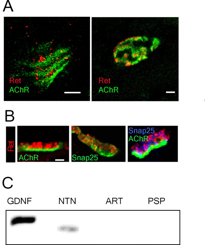
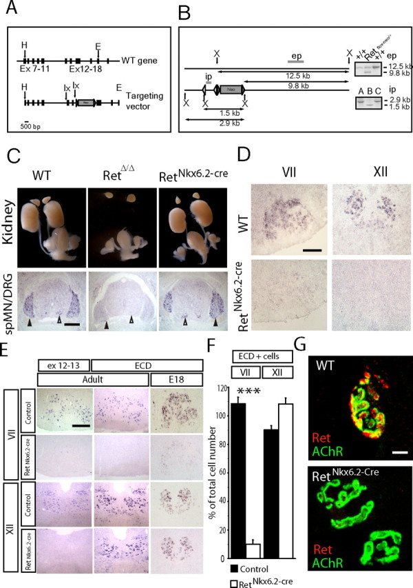
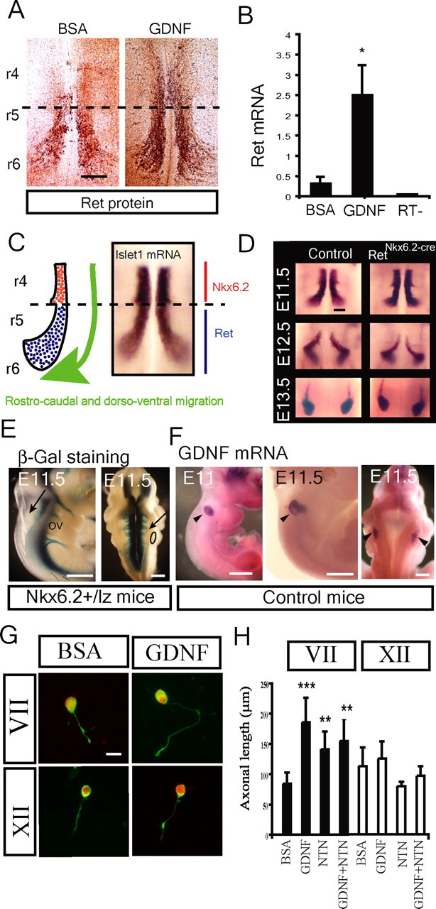
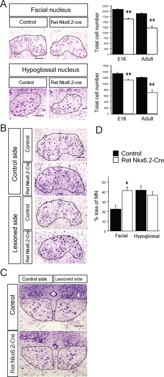
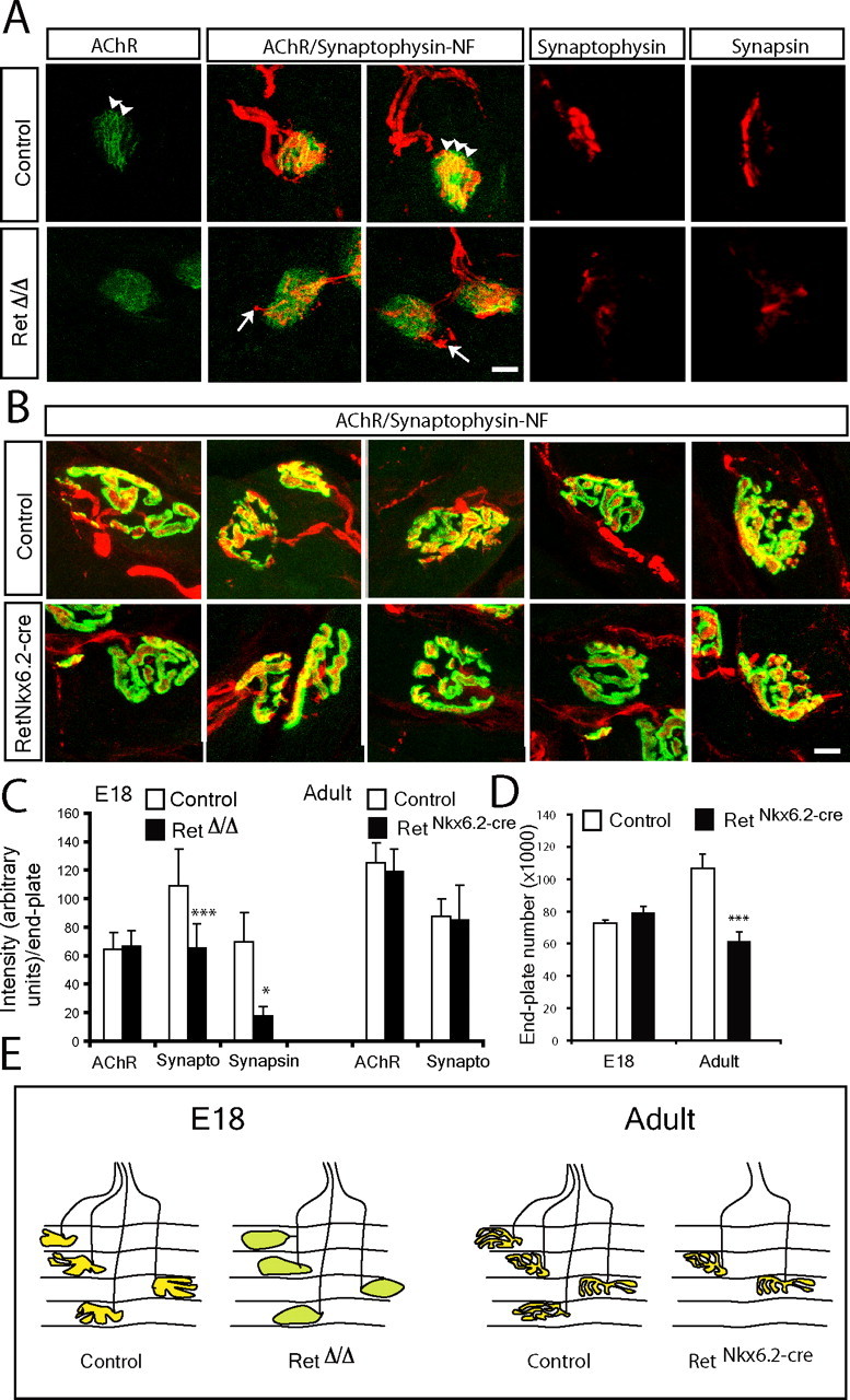
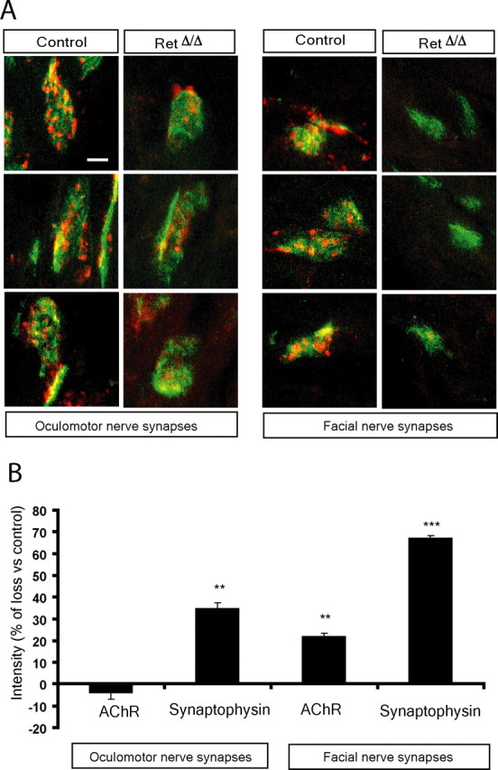
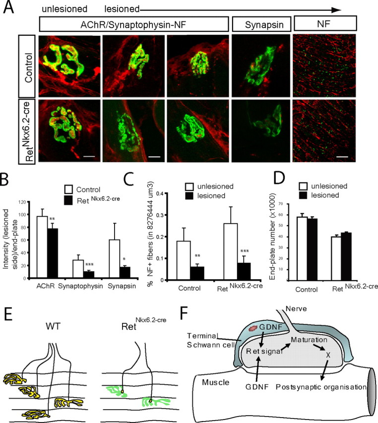
Establishment of the neuromuscular synapse requires bidirectional signaling between the nerve and muscle. Although much is known on nerve-released signals onto the muscle, less is known of signals important for presynaptic maturation of the nerve terminal. Our results suggest that the Ret tyrosine kinase receptor transmits a signal in motor neuron synapses that contribute to motor neuron survival and synapse maturation at postnatal stages. Ret is localized specifically to the presynaptic membrane with its ligands, GDNF (glial cell line-derived neurotrophic factor)/NTN (neurturin), expressed in skeletal muscle tissue. Lack of Ret conditionally in cranial motor neurons results in a developmental deficit of maturation and specialization of presynaptic neuromuscular terminals. Regeneration of Ret-deficient adult hypoglossal motor neurons is unperturbed, but despite contact with the unaffected postsynaptic specializations, presynaptic axon terminal maturation is severely compromised in the absence of Ret signaling. Thus, Ret transmits a signal in motor nerve terminals that participate in the organization and maturation of presynaptic specializations during development and during regeneration in the adult.


Figures

Figure 1.
Figure 1.
Concentration of Ret receptors presynaptically in the neuromuscular synapse and expression of GDNF family ligands in the target tissue. A, Double labeling for Ret and AChRs (bungarotoxin binding) on the internal muscle of the tongue shows the presence of Ret in the neuromuscular synapse of E18 and adult mice. B, Confocal images show the specific presence of Ret receptors in the presynaptic area (labeled by Snap-25 immunohistochemistry) but not in the postsynaptic membrane (stained by bungarotoxin binding to AChRs). C, RT-PCR for all GDNF family ligands shows the expression of GDNF and NTN in the neonatal tongue. Scale bars, 4 μm.
Figure 2.
Figure 2.
Conditional inactivation of Ret in cranial motor neurons. A, Strategy for producing a conditional Ret allele with schematic representation of the wild-type Ret locus and the targeting vector. The black rectangles represent exons, and the gray triangles represent Frt sites. LoxP (lx) sites are indicated by arrows. The targeting vector, a 12 kb HindIII(H)/EcoRI(E) fragment from the mouse Ret gene contains exons 7–18. One loxP site was introduced in intron 11. The second loxP, inserted in intron 13, was directly followed by the selection cassette (Neo) flanked by Frt sites. B, Schematic representation of the strategy used to analyze homologous recombination by Southern blot and select the ES cell clones (Retflox-neo/+) for blastocyst injection. Digestion with XbaI (X) generated a 12.5 kb band for the wild-type allele and a 9.8 kb band for the floxed allele using the external probe (ep) probe. The internal probe (ip) was used to discriminate ES cell clones in which the homologous recombination had occurred between the LoxP sites; expected sizes are indicated. Flp- and Cre-mediated excision was used to produce RetNkx6.2-Cre and RetΔ/Δ animals. RetΔ/Δ mice carry a deletion of exons 12 and 13 in all cells (germline mutation), whereas RetNkx6.2-Cre mice carry this deletion only in cells expressing Nkx6.2. C, Genetic excision of Ret exons 12 and 13 analyzed in E18 embryos. RetΔ/Δ mice carrying a deletion of Ret in the germline displayed kidney agenesis and absence of Ret exons 12 and 13 expression in spinal cord motor neurons and dorsal root ganglion by in situ hybridization. RetNkx6.2-Cre mice show an intact urogenital system and kidney, and Ret expression can be detected in spinal cord motor neurons (spMNs) (white arrowheads) and in the dorsal root ganglia (DRG) (black arrowheads). D, RetNkx6.2-Cre mice show a specific loss of Ret exons 12 and 13 expression in the facial (VII) and hypoglossal (XII) MNs. E, F, Motor neuron-specific regulation of Ret expression in cranial nuclei VII and XII. E, In situ hybridization using the ex 12–13 and ECD probes on coronal section of the brainstem for the indicated motor nuclei, ages, and genotypes. F, Ret ECD expression in motor neurons as percentage of total cell number in adult animals. Note the dramatic and specific loss of Ret transcript expression in the facial (VII) but not hypoglossal (XII) nucleus. G, Complete loss of Ret protein at the neuromuscular synapse of RetNkx6.2-Cre mice (n = 4–6 nuclei; ***p < 0.0001). Scale bars: C, 250 μm; D, E, 100 μm; G, 5 μm.
Figure 3.
Figure 3.
Ret activated Ret expression and Ret induced axonal growth in facial but not hypoglossal motor neurons. A, GDNF (50 ng/ml) upregulated Ret protein expression in organotypic culture of E11.5 hindbrains [rhombomere 4–6 (r4–r6)] after 6 h compared with BSA-treated control. Whole-mount immunostaining with Ret antibody on the control samples shows Ret expression restricted to migratory and postmigratory motoneurons (r5–r6, limited by dashed lines). GDNF induces increased expression of Ret in migrating motor neurons (r5–r6) as well as a precocious expression by premigatory motoneurons (r4). B, GDNF upregulated Ret mRNA expression in organotypic culture of E11.5 hindbrains [rhombomere 4–6 (r4–r6)] after 6 h compared with BSA-treated control (RT−, reaction without reverse transcriptase). Quantitative real-time PCR was conducted for ret mRNA (mean ± SEM of 3 independent experiments). C, Schematic illustration and islet1 in situ hybridization of an open-book preparation of hindbrain showing the path of facial motor neuron migration. Premigratory (red) cells express Nkx6.2, whereas migrating and postmigratory cells express Ret (blue) (Vallstedt et al., 2001). D, Islet1 staining revealing that elimination of Ret as detected in RetNkx6.2-Cre mice did not affect cell migration. E–H, GDNF is expressed along the axonal pathway of the facial nerve and promotes axonal growth of facial but not hypoglossal motoneurons in primary culture. E, The facial nerve exits the hindbrain at the level of the otic vesicle (arrows) as shown by the lateral (left) and dorsal (right) view of E11.5 Nkx6.2+/lz embryos after β-gal staining. F, Specific expression of GDNF mRNA in the otic vesicle of wild-type E11 and E11.5 embryos (black arrowheads) as shown by in situ hybridization. G, H, Axonal growth of facial but not hypoglossal motor neurons is affected by GDNF and NTN. G, Facial and hypoglossal motoneurons from E11.5 wild-type embryos were cultured in the presence of BSA or 50 ng/ml GDNF and stained for peripherin (green) and Islet1 (red) and the length of the axons measured. H, Quantification of axonal length in the presence of BSA or 50 ng/ml neurotrophic factors (GDNF, NTN, and GDNF plus NTN) in primary culture of facial and hypoglossal motor neurons (n = 50–100). Graphs represent the mean ± SEM. *p ≤ 0.05; **p ≤ 0.005; ***p ≤ 0.0005. Scale bars: A, 100 μm; E, F, 250 μm; H, 20 μm.
Figure 4.
Figure 4.
Postnatal motor neuron death in the absence of Ret. A, Motor neuron loss in facial and hypoglossal motor nuclei of adult Ret-deficient mice. Cresyl violet staining of coronal sections of facial and hypoglossal nuclei and graph showing the total number of motoneurons in control (black bars; n = 5–12 nuclei) and RetNkx6.2-Cre mice (white bars; n = 4–14 nuclei) at E18 and 3–4 months (adult). Nuclei are outlined by the dashed lines. Graphs represent the mean ± SEM. All the groups were statistically significant from controls; **p ≤ 0.005. B–D, Ret inactivation decreases the survival of facial but not hypoglossal adult motor neurons 21 d after nerve lesion. B, Photomicrographs of cresyl violet staining of the right facial nucleus (control side) and the left facial nucleus (lesioned side) from control and RetNkx6.2-Cre mice, as indicated. The dashed line represents the limit of the nucleus. C, Photomicrographs of sections showing the right hypoglossal nucleus (control side) and the left hypoglossal nucleus (lesioned side) from control and RetNkx6.2-Cre mice, as indicated. The dashed lines delimit the nucleus. D, Quantification of facial and hypoglossal motor neuron loss by the lesion as percentage of the control side in control mice (control, n = 3 mice; RetNkx6.2-Cre, n = 9 mice). Note the significant loss of facial but not hypoglossal MN as a consequence of the Ret genomic inactivation (*p < 0.015). Scale bars, 100 μm.
Figure 5.
Figure 5.
Deficits of synapse maturation in the absence of Ret signaling. A, B, Photomicrographs of neuromuscular synapses in the E18 (A) and adult (B) tongue of control and the Ret-deficient mice (RetΔ/Δ in A and RetNkx6.2-Cre in B). The presynaptic region was stained by synapsin I or synaptophysin immunohistochemistry (red, as indicated). AChRs in the postsynaptic membrane were labeled by fluorescent coupled bungarotoxin binding (green). Neuromuscular junctions visualized by a triple staining that combined synaptophysin-neurofilament (synaptophysin-NF; red) with bungarotoxin binding (AChR; green) are indicated. In A, note the reduction in the immunostaining of presynaptic components (synapsin I and synaptophysin) in Ret-deficient mice. The postsynaptic AChRs are more aggregated in control than RetΔ/Δ mice at E18 (arrowheads). Also note terminal sprouts in RetΔ/Δ mice (arrows in A), whereas general morphology of both presynaptic and postsynaptic elements of the neuromuscular junction is similar between adult control and RetNkx6.2-Cre mice (B). C, Quantification of synaptophysin (synapto) and synapsin immunoreactivity as a measure of synapse maturations at the neuromuscular synapse and AChRs staining intensity at E18 and in adult mice. Note the significant loss of synaptophysin and synapsin but not postsynaptic AChRs in the absence of Ret signaling at E18. No significant difference was found in adult mice. D, Quantification of the total number of AChR containing endplates in serial sections through the entire tongue at E18 and in the adult of both genotypes. Note a significant reduction of the total number of endplates in adult RetNkx6.2-Cre mice. E, Schematic illustration summarizing our interpretation of the results from the above studies depicting vesicle containing presynaptic terminals overlaying the postsynaptic AChR membrane in control mice (yellow). In E18 RetΔ/Δ mice, synapses are less developed (green) but in similar number as in control embryos. Adult RetNkx6.2-Cre mice display a reduced number of synapses, which otherwise appear normal. Graphs represent the mean ± SEM. ***p ≤ 0.005; *p ≤ 0.05. Scale bars, 10 μm.
Figure 6.
Figure 6.
Developmental deficit of synapse maturation in the absence of Ret signaling in facial and oculomotor nerve synapses. A, Photomicrographs of neuromuscular synapses in the E18 facial and oculomotor and muscles. The nerve and presynaptic region was stained by synaptophysin and neurofilament immunohistochemistry (red) and AChRs in the postsynaptic membrane by fluorescent-coupled bungarotoxin binding (green). Note similar deficits of synapse maturation in these muscles as in the tongue of RetΔ/Δ mice. B, Quantification of AChR and synaptophysin immunoreactivity at the neuromuscular synapse. Note deficits in synaptophysin immunoreactivity in RetΔ/Δ mice of both muscles, with a more pronounced deficit in the facial nerve synapses. Also note loss of AChR in the buccinator but not oculomotor muscle. Graphs represent the mean ± SEM. *p ≤ 0.05; **p ≤ 0.005; ***p ≤ 0.0005. Scale bar, 10 μm.
Figure 7.
Figure 7.
Requirement of Ret for maturation of regenerating neuromuscular synapses in the adult mouse. A–D, The hypoglossal nerve was lesioned and reinnervation of synaptic sites at the muscle examined 3 weeks later in the muscle. A, Immunohistochemistry for neurofilament (NF) and synaptophysin immunohistochemistry (red) or synapsin I (red, where indicated) and AChRs (green), comparing tongue sections from the side where the hypoglossal nerve was lesioned and the contralateral unlesioned side, as indicated. After lesion, the neurofilament immunostaining shows that reinnervation occurs in the tongue of both control and mutant mice, but the establishment of the mature synaptic contact after reinnervation detected in control is absent in RetNkx6.2-Cre mice. B, Quantification of AChRs, synaptophysin, and synapsin I intensity in control and reinnerverated muscles of RetNkx6.2-Cre mice versus control mice shows a strong reduction of all measured intensities. C, Quantification neurofilament staining in a defined volume of each side of the tongue (unlesioned vs lesioned) for control and mutant mice. Note the reduction of NF staining on the lesioned site of the tongue both in control and RetNkx6.2-Cre mice. There was no statistical significance between control and RetNkx6.2-Cre mice. D, The number of postsynaptic endplates in one-half of the tongue (lesioned and unlesioned sides, as indicated) of control and RetNkx6.2-Cre mice. Note that the number of postsynaptic structures were not affected by Ret deletion. E, Schematic illustration summarizing the results from the above studies. Although we find mature presynaptic terminals localizing with AChR-rich regions (yellow) in control animals 3 weeks after lesion, RetNkx6.2-Cre mice show only punctuate presynaptic contact in all endplates (yellow) overlaying AChR postsynaptic organizations (green), but displaying severe deficits of presynaptic maturation. F, Proposed model for Ret signaling in the establishment and maturation of the neuromuscular synapse. The solid arrows indicate processes with direct or indirect supporting data, and the dashed arrow indicates a suggestion that postsynaptic maturation may affect GDNF production/release and thereby also affect presynaptic maturation. **p ≤ 0.005; ***p ≤ 0.0005. Scale bars: NF, 100 μm; all other images, 10 μm.

References

    1. Abercrombie M. Estimation of nuclear population from microtome sections. Anat Rec. 1946;94:239–247. - PubMed
    1. Balice-Gordon RJ, Lichtman JW. In vivo visualization of the growth of pre- and postsynaptic elements of neuromuscular junctions in the mouse. J Neurosci. 1990;10:894–908. - PMC - PubMed
    1. Balice-Gordon RJ, Lichtman JW. In vivo observations of pre- and postsynaptic changes during the transition from multiple to single innervation at developing neuromuscular junctions. J Neurosci. 1993;13:834–855. - PMC - PubMed
    1. Baudet C, Mikaels A, Westphal H, Johansen J, Johansen TE, Ernfors P. Positive and negative interactions of GDNF, NTN and ART in developing sensory neuron subpopulations, and their collaboration with neurotrophins. Development. 2000;127:4335–4344. - PubMed
    1. Bibel M, Barde YA. Neurotrophins: key regulators of cell fate and cell shape in the vertebrate nervous system. Genes Dev. 2000;14:2919–2937. - PubMed

Publication types

Substances

LinkOut - more resources