. 2008 Jan 29;105(4):1291-6.
doi: 10.1073/pnas.0711257105. Epub 2008 Jan 23.

The insulin-like growth factor pathway is altered in spinocerebellar ataxia type 1 and type 7

Affiliations

The insulin-like growth factor pathway is altered in spinocerebellar ataxia type 1 and type 7

Jennifer R Gatchel et al. Proc Natl Acad Sci U S A. .

Abstract

Polyglutamine diseases are inherited neurodegenerative disorders caused by expansion of CAG repeats encoding a glutamine tract in the disease-causing proteins. There are nine disorders, each having distinct features but also clinical and pathological similarities. In particular, spinocerebellar ataxia type 1 and 7 (SCA1 and SCA7) patients manifest cerebellar ataxia with degeneration of Purkinje cells. To determine whether the disorders share molecular pathogenic events, we studied two mouse models of SCA1 and SCA7 that express the glutamine-expanded protein from the respective endogenous loci. We found common transcriptional changes, with down-regulation of insulin-like growth factor binding protein 5 (Igfbp5) representing one of the most robust changes. Igfbp5 down-regulation occurred in granule neurons through a non-cell-autonomous mechanism and was concomitant with activation of the insulin-like growth factor (IGF) pathway and the type I IGF receptor on Purkinje cells. These data define one common pathogenic response in SCA1 and SCA7 and reveal the importance of intercellular mechanisms in their pathogenesis.


Conflict of interest statement

The authors declare no conflict of interest.

Figures
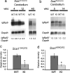
Fig. 1.
Fig. 1.
Igfbp5 RNA is decreased in Sca1154Q/2Q and Sca7266Q/5Q cerebellum. (a and b) Northern blots of cerebellar RNA at early-symptomatic (Sca1154Q/2Q, 4 and 12 weeks; Sca7266Q/5Q, 5 weeks) and late-symptomatic (Sca1154Q/2Q, 40 weeks; Sca7266Q/5Q, 14 weeks) disease stages show progressive down-regulation of Igfbp5 RNA. (c and d) Quantification of Igfbp5 RNA relative to Gapdh using multiple animal pairs (n = 3 pairs of Sca1154Q/2Q and WT controls; n = 5 and 4 of Sca7266Q/5Q and WT mice, respectively) shows significant down-regulation of Igfbp5 in late-symptomatic cerebellum. Values represent mean ± SEM. The asterisks indicate significant genotype differences (*, P < 0.05).
Fig. 2.
Fig. 2.
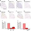
Igfbp5 is decreased in the cerebellar granule layer of Sca1 and Sca7 KI mice and in SCA1[82Q] PC-specific Tg mice. (a–h) Representative ISH images for Igfbp5 (a, c, e, and g) and pseudocolored sagittal sections (b, d, f, and h) with Igfbp5-expressing cells colored according to their expression level (red, strong; blue, moderate; yellow, weak). (i) Quantification of strongly and moderately expressing cells reveals a decrease in Sca1154Q/2Q (18–19 weeks) and Sca7266Q/5Q (12–13 weeks) cerebellum compared with WT. A decrease is also observed when mutant ATXN1 is only expressed in PC, in SCA1[82Q]Tg/+ (22–23 weeks) cerebellum. n = 2 KI and 2 WT animals for both Sca1154Q/2Q and Sca7266Q/5Q and 2 Tg/+ and 2 WT littermates for SCA1[82Q], with 40–47 sections quantified per animal. KI and Tg/+ expression values are shown as a percentage of WT. Values represent mean ± SEM.
Fig. 3.
Fig. 3.
IGF1R phosphorylation is increased in Sca1154Q/2Q, Sca7266Q/5Q, and SCA1[82Q] Tg cerebellum. (a, c, and e) Western blot analyses using cerebellar protein extracts from Sca1154Q/2Q (49–52 weeks) and Sca7266Q/5Q (12–13 weeks) mice and SCA1[82Q] (8–10 weeks) mice and antibodies to the phosphorylated (active) form of the IGF1R as well as to Gapdh. (b, d, and f) Quantification of phospho-IGF1R levels relative to Gapdh using multiple pairs of KI, Tg, and WT animals (n = 3; see Methods) indicates a significant increase in Sca1 KI and Sca7 KI vs. WT (b and d) as well as a significant increase in SCA1[82Q]Tg/Tg and Tg/+ compared with WT (f). Values represent mean ± SEM. The asterisks indicate significant genotype differences (*, P < 0.05; **, P < 0.01).
Fig. 4.
Fig. 4.
Patterns of IGF1R and ERK1/2 phosphorylation relative to PC pathology in SCA1[82Q]Tg/Tg cerebellum. (a) Representative confocal images from 11-week-old SCA1[82Q]Tg/Tg and WT mice showing calbindin and phospho-IGF1R immunostaining in sections from anterior cerebellar lobules and from lobule 10. (b) Calbindin immunostaining is decreased, and phospho-IGF1R levels are increased in the PC, molecular, and granule layers in Tg/Tg cerebellum compared with WT. Immunoreactivity for phosphorylated ERK, which is increased in SCA1[82Q]Tg/Tg cerebellum (SI Fig. 24a), occurs most prominently in anterior lobules compared with lobule 10. Similar results were obtained from three pairs of SCA1[82Q]Tg/Tg and WT mice (three to four sections per animal) in four independent experiments. (Scale bars, 100 μm.)
Fig. 5.
Fig. 5.
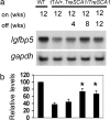
Igfbp5 down-regulation is reversible in an SCA1[82Q] conditional mouse model. (a) Northern blot analysis shows the gradual recovery of Igfbp5 RNA levels after the mutant SCA1 transgene has been turned off for 4, 8, or 12 weeks. (b) Igfbp5 RNA levels relative to Gapdh in SCA1[82Q] cerebellum were normalized to WT, with three animals in each group. Data are expressed as mean ± SEM; (*, P < 0.05).
Fig. 6.
Fig. 6.
Model of Igfbp5 and IGF1 signaling in normal (A) and in Sca1154Q/2Q, Sca7266Q/5Q, and SCA1[82Q] (B) cerebellum. Igfbp5 is expressed in granule neurons, whereas the IGF1R is more broadly expressed, suggesting possible cross-talk between PCs and granule neurons. In normal cerebellum, IGF1 can be bound by Igfbp5, which influences its availability for the IGF1R (A). Mutant ATXN1- and ATXN7-induced PC injury causes down-regulation of Igfbp5 in granule neurons by a non-cell-autonomous mechanism (B). This mechanism may involve IGF1, expressed in part by PCs or, alternatively, could result from granule neurons sensing altered synaptic activity at granule–PC–parallel fiber connections. Given the increased IGF1R activation in Sca1154Q/2Q, Sca7266Q/5Q, and SCA1[82Q] cerebellum, we propose a model in which down-regulation of Igfbp5 in granule neurons influences the local actions of IGF1, in part by increasing the availability of IGF1 for its receptor on PCs, which can lead to activation of downstream effectors such as Akt.

References

    1. Orr HT, Zoghbi HY. Trinucleotide repeat disorders. Annu Rev Neurosci. 2007;30:575–621. - PubMed
    1. Borrell-Pages M, Zala D, Humbert S, Saudou F. Huntington's disease: From huntingtin function and dysfunction to therapeutic strategies. Cell Mol Life Sci. 2006;63:2642–2660. - PMC - PubMed
    1. Stevanin G, et al. Spinocerebellar ataxia type 7. In: Hayden MR, Rubinsztein DC, editors. Analysis of Triplet Repeat Disorders. Oxford, UK: BIOS Scientific; 1998. pp. 155–168.
    1. Tsai CC, et al. Ataxin 1, a SCA1 neurodegenerative disorder protein, is functionally linked to the silencing mediator of retinoid and thyroid hormone receptors. Proc Natl Acad Sci USA. 2004;101:4047–4052. - PMC - PubMed
    1. Serra HG, et al. RORalpha-mediated Purkinje cell development determines disease severity in adult SCA1 mice. Cell. 2006;127:697–708. - PubMed

Publication types

MeSH terms