. 2008 Apr;19(4):1354-65.
doi: 10.1091/mbc.e07-11-1110. Epub 2008 Jan 23.

UME6, a novel filament-specific regulator of Candida albicans hyphal extension and virulence

Affiliations

UME6, a novel filament-specific regulator of Candida albicans hyphal extension and virulence

Mohua Banerjee et al. Mol Biol Cell. 2008 Apr.

Abstract

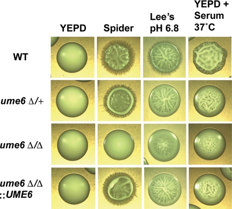
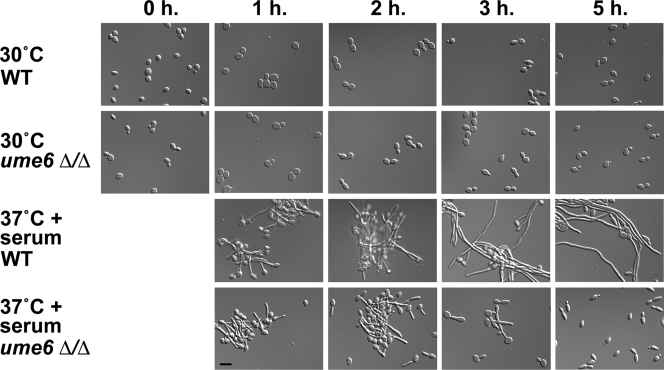
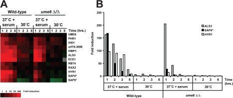
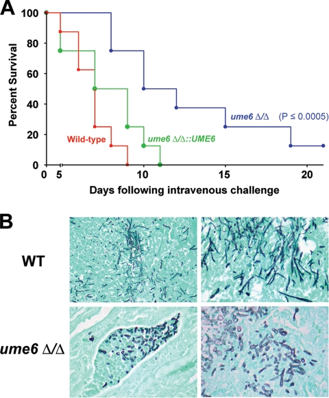
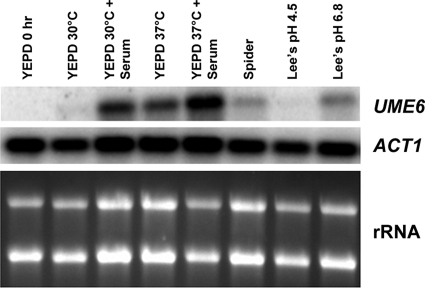
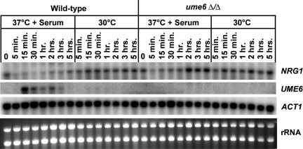
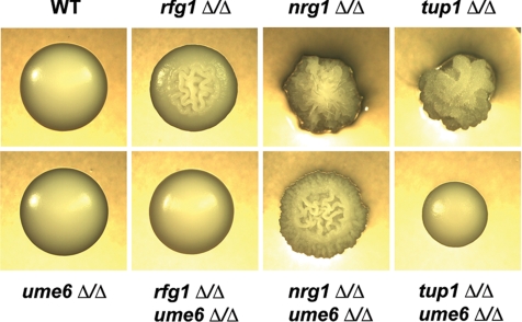
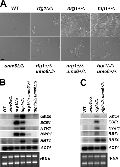
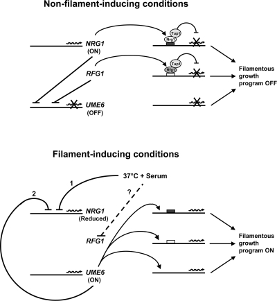
The specific ability of the major human fungal pathogen Candida albicans, as well as many other pathogenic fungi, to extend initial short filaments (germ tubes) into elongated hyphal filaments is important for a variety of virulence-related processes. However, the molecular mechanisms that control hyphal extension have remained poorly understood for many years. We report the identification of a novel C. albicans transcriptional regulator, UME6, which is induced in response to multiple host environmental cues and is specifically important for hyphal extension. Although capable of forming germ tubes, the ume6Delta/ume6Delta mutant exhibits a clear defect in hyphal extension both in vitro and during infection in vivo and is attenuated for virulence in a mouse model of systemic candidiasis. We also show that UME6 is an important downstream component of both the RFG1-TUP1 and NRG1-TUP1 filamentous growth regulatory pathways, and we provide evidence to suggest that Nrg1 and Ume6 function together by a negative feedback loop to control the level and duration of filament-specific gene expression in response to inducing conditions. Our results suggest that hyphal extension is controlled by a specific transcriptional regulatory mechanism and is correlated with the maintenance of high-level expression of genes in the C. albicans filamentous growth program.


Figures

Figure 1.
Figure 1.
ume6Δ/Δ mutants are defective for filamentous growth in response to a variety of filament-inducing conditions. Colony morphologies of wild-type (WT), ume6Δ/+, ume6Δ/Δ, and ume6Δ/Δ::UME6 (add-back) strains grown on solid non–filament-inducing medium (YEPD) and on various solid filament-inducing media. All colonies were grown at 30°C for 5 d, with the exception of cells grown on YEPD + 10% serum plates, which were grown at 37°C for 3 d, and photographed at ∼×20 magnification.
Figure 2.
Figure 2.
Morphology of wild-type and ume6Δ/Δ cells undergoing the blastospore to filament transition. Wild-type (WT) and ume6Δ/Δ strains grown under non–filament-inducing conditions (YEPD medium at 30°C) were diluted into pre-warmed YEPD medium at 30°C or 37°C in the presence or absence of 10% fetal calf serum (FCS). Induction time (h) is shown on top. Aliquots of cells were fixed in 4.5% formaldehyde, washed twice in 1× PBS, and then visualized by Nomarski/DIC optics. Please note that the 0-h time point shows cells immediately before induction. Bar, 10 μm.
Figure 3.
Figure 3.
Transcriptional profile of serum- and temperature-induced genes in wild-type and ume6Δ/Δ strains. (A) Cluster diagram indicating expression levels of several previously identified (Kadosh and Johnson, 2005) top serum- and temperature-induced transcripts (≥5-fold mean induction in the wild-type strain, n = 2, at the 37°C + 10% FCS 1-h time point). Only data from one serum- and temperature-induction experiment and only genes with greater than 93% of data present are shown. Red, increased expression; green, reduced expression; gray, no data available. Expression levels reflect fold induction relative to the zero time point for each strain. None of the genes showed a significant difference in absolute level of expression between wild-type and ume6Δ/Δ strains at the zero time point. (B) Histogram indicates fold induction, relative to the zero time point of wild-type or ume6Δ/Δ strains, for three serum- and temperature-induced genes: ALS3, an adhesin important for both epithelial and endothelial cell adhesion (Zhao et al., 2004), SAP4, a secreted aspartyl protease important for host tissue invasion and virulence (Sanglard et al., 1997; Schaller et al., 1999), and HYR1, a putative cell wall glycoprotein (Bailey et al., 1996) described in A. *Note that expression levels of SAP5 may, in part, reflect those of SAP4 or SAP6 (and SAP4 levels may reflect SAP5 and SAP6 levels) due to cross-hybridization on the microarray, since these genes have nearly identical DNA sequences.
Figure 4.
Figure 4.
The ume6Δ/Δ mutant is attenuated for virulence and defective for hyphal filament extension in a mouse model of systemic candidiasis. (A) Eight female BALB/c mice (6–8 wk old) were each injected with 2 × 10⁵ CFUs of a wild-type, ume6Δ/Δ, or ume6Δ/Δ::UME6 (add-back) strain. Survival was monitored over the course of 21 d. A Kaplan-Meier test was performed to confirm that the difference in virulence between the wild-type and ume6Δ/Δ strains is statistically significant (p ≤ 0.0005) (the ume6Δ/Δ::UME6 strain did not show a statistically significant difference in virulence when compared with the wild-type strain). (B) Kidney tissues from infected mice were fixed, sectioned, and stained with Grocot- Gomori methenamine-silver to visualize fungal cells. Top panels, sections taken from mice infected with the wild-type (WT) strain; bottom panels, sections taken from mice infected with the ume6Δ/Δ strain.
Figure 5.
Figure 5.
Effect of a variety of filament-inducing conditions on induction of UME6. A wild-type strain was grown under non–filament-inducing conditions (YEPD medium at 30°C) and diluted into pre-warmed flasks containing either YEPD or the indicated filament-inducing media at 30°C (or 37°C where shown). Cells were harvested at the zero time point (immediately before induction) and at 30 min after induction for total RNA preparation. Northern analysis was carried out using 3 μg of RNA from each sample to assess transcript levels of the indicated genes. The ACT1 transcript, as well as ribosomal RNA (rRNA) are shown as loading controls.
Figure 6.
Figure 6.
Kinetics of NRG1 down-regulation and UME6 induction in response to serum at 37°C. Wild-type and ume6Δ/Δ strains were grown under non–filament-inducing conditions (YEPD medium at 30°C) and diluted into pre-warmed YEPD medium at 30°C or YEPD medium + 10% FCS at 37°C. Cells were harvested at the zero time point (immediately before induction) and at the indicated time points after induction for total RNA preparation. Northern analysis was carried out using 3 μg of RNA from each sample to assess transcript levels of the indicated genes. The ACT1 transcript, as well as ribosomal RNA (rRNA) are shown as loading controls.
Figure 7.
Figure 7.
Colony morphologies of rfg1Δ/Δ ume6Δ/Δ, nrg1Δ/Δ ume6Δ/Δ, and tup1Δ/Δ ume6 Δ/Δ double mutants and respective single mutants. Colony morphologies of the indicated strains are shown after growth on solid YEPD medium at 30°C (non–filament-inducing conditions) for 3 d. Colonies were visualized by light microscopy and photographed at ∼×20 magnification.
Figure 8.
Figure 8.
Cell morphologies and filament-specific gene expression in rfg1Δ/Δ ume6Δ/Δ, nrg1Δ/Δ ume6Δ/Δ, and tup1Δ/Δ ume6Δ/Δ double mutants and respective single mutants. (A) Cell morphologies of the indicated strains are shown after growth in liquid YEPD medium at 30°C to an OD600 ∼ 1.0. Aliquots of cells were fixed in 4.5% formaldehyde, washed twice in 1× PBS, and then visualized by Nomarski/DIC optics (Bar, 10 μm). (B and C) Total RNA was prepared from the strains shown in A, and Northern analysis was carried using 3 μg RNA and probes to the indicated filament-specific transcripts. The ACT1 transcript and ribosomal RNA (rRNA) are included as loading controls.
Figure 9.
Figure 9.
Model for control of induction and maintenance of expression of the C. albicans filamentous growth program by Rfg1, Nrg1, Tup1, and Ume6. Nrg1 and Ume6 are believed to repress each other in a negative feedback loop. Under steady-state non–filament-inducing conditions (top) Nrg1 levels are high, and UME6 is not expressed (due to strong Nrg1-Tup1 repression). In turn, many filament-specific transcripts are not induced as a consequence of repression by the Nrg1-Tup1 pathway and/or lack of activation by Ume6. In addition, under non–filament-inducing conditions, UME6 as well as several filament-specific transcripts, are repressed by the Rfg1-Tup1 pathway. In the presence of filament-inducing conditions such as serum at 37°C (bottom) the NRG1 transcript undergoes a very rapid transient down-regulation (1) causing UME6 levels to rise. As a consequence of repression by Ume6 (2), the NRG1 transcript is maintained at a reduced level for a significantly longer time period. Filament-specific transcripts are subsequently induced as a result of relief of repression by the Nrg1-Tup1 and Rfg1-Tup1 pathways and/or activation by Ume6. Also, in the presence of serum at 37°C repression of UME6 by the Rfg1-Tup1 pathway is believed to be relieved by a mechanism not yet determined (dashed line). Please note that it is unclear at this point as to whether Ume6 and Nrg1 repress each other by direct or indirect effects.

References

    1. Arnaud M. B., Costanzo M. C., Skrzypek M. S., Binkley G., Lane C., Miyasato S. R., Sherlock G. The Candida Genome Database (CGD), a community resource for Candida albicans gene and protein information. Nucleic Acids Res. 2005;33:D358–D363. - PMC - PubMed
    1. Ausubel F. M., Brent R., Kingston R. E., Moore D. D., Seidman J. G., Smith J. A., Struhl K., editors. New York: Greene Publishing Associates and Wiley-Interscience; 1992. Current Protocols in Molecular Biology.
    1. Bailey D. A., Feldmann P. J., Bovey M., Gow N. A., Brown A. J. The Candida albicans HYR1 gene, which is activated in response to hyphal development, belongs to a gene family encoding yeast cell wall proteins. J. Bacteriol. 1996;178:5353–5360. - PMC - PubMed
    1. Bartie K. L., Williams D. W., Wilson M. J., Potts A. J., Lewis M. A. Differential invasion of Candida albicans isolates in an in vitro model of oral candidosis. Oral Microbiol. Immunol. 2004;19:293–296. - PubMed
    1. Bennett R. J., Uhl M. A., Miller M. G., Johnson A. D. Identification and characterization of a Candida albicans mating pheromone. Mol. Cell. Biol. 2003;23:8189–8201. - PMC - PubMed

Publication types

MeSH terms

LinkOut - more resources