Review
. 2008 Jan 23;27(2):315-27.
doi: 10.1038/sj.emboj.7601974.

Protein quality control in the early secretory pathway

Affiliations
Review

Protein quality control in the early secretory pathway

Tiziana Anelli et al. EMBO J. .

Abstract

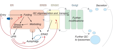
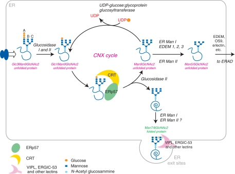
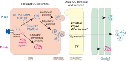
Eukaryotic cells are able to discriminate between native and non-native polypeptides, selectively transporting the former to their final destinations. Secretory proteins are scrutinized at the endoplasmic reticulum (ER)-Golgi interface. Recent findings reveal novel features of the underlying molecular mechanisms, with several chaperone networks cooperating in assisting the maturation of complex proteins and being selectively induced to match changing synthetic demands. 'Public' and 'private' chaperones, some of which enriched in specializes subregions, operate for most or selected substrates, respectively. Moreover, sequential checkpoints are distributed along the early secretory pathway, allowing efficiency and fidelity in protein secretion.


Figures

Figure 1
Figure 1
The early secretory pathway. Proteins destined to the extracellular space or to organelles of the secretory route are synthesized by ER-bound ribosomes and cotranslationally translocated (entry) into the ER. Here they attain their native structure (folding), under strict QC scrutiny. Only properly folded and assembled proteins can reach the Golgi, where they are further modified, to be transported to the extracellular space or to lysosomes. Gray arrows indicate the direction of vesicles moving among different compartments; dark arrows indicate the pathways followed by cargoes in the early secretory pathway; red lines show homeostatic control pathways (+ stimulatory, − inhibitory). Misfolded proteins are recognized, retained and eventually routed to degradation by ERAD or autophagy (which are likely reciprocally regulated, as indicated by the blue arrows). Some misfolded soluble ERAD substrates are transported to ERGIC or cis-Golgi before retrotranslocation and degradation. Too high load for the folding machinery or the accumulation of misfolded proteins activate resident ER stress sensors, which elicit the UPR. ER stress can selectively inhibit protein entry into the ER, and increase the capacity of folding and degradation (via ERAD and autophagy). The UPR induces also molecules acting downstream of the ER.
Figure 2
Figure 2
The CNX/CRT cycle. After transfer of the preformed core oligosaccharide (Glc3Man9GlcNAc2) onto nascent proteins, glucosidase I and II sequentially remove the two terminal glucoses from the A branch. The monoglucosylated Glc1Man9GlcNAc2 unfolded protein can now interact with the lectin chaperones CNX and CRT. In association with the oxidoreductase ERp57, CNX and CRT prevent aggregation and facilitate glycoprotein folding. Removal of the glucose by glucosidase II (Man9GlcNAc2) interrupts the interaction of the protein with CNX/CRT. If the protein has attained its native structure, it can now proceed along the secretory pathway by bulk flow or by interaction with specific lectin transporters such as ERGIC-53 or VIPL. If unfolding persists, the glycoprotein is recognized by UGGT1, which places a single glucose back onto the A branch, causing the protein to enter the CNX/CRT cycle again. Mannose trimming causes exit from the CNX/CRT cycle. Misfolded proteins can be recognized by specific lectins (EDEMs, OS9, etc) and targeted to degradation.
Figure 3
Figure 3
QC in the early secretory pathway. Chaperones and folding assistants can be grouped in different classes according to their specificity and subcellular localization. The majority of secretory proteins utilize public chaperones: some initially go with BiP, PDI and their partners, others enter the CNX/CRT cycle, the choice depending on the location of the N-glycans. Certain proteins that are produced in large amounts, or are intrinsically difficult to fold, are assisted by specific (private) chaperones and enzymes (see also Box 2). In addition, QC can occur in sequential steps. After a proximal QC, certain substrates (generally multimeric proteins) seem to undergo also distal QC checkpoints in ERGIC and cis-Golgi. This model could mediate cargo concentration and selective export of oligomerized species, thus coupling fidelity and efficiency in the secretory protein factory. While proximal QC can rely on simple retention, the distal checkpoints likely imply substrate retrieval to the ER, either for further attempts to fold, or for retrotranslocation and degradation.

References

    1. Ahluwalia N, Bergeron JJ, Wada I, Degen E, Williams DB (1992) The p88 molecular chaperone is identical to the endoplasmic reticulum membrane protein, calnexin. J Biol Chem 267: 10914–10918 - PubMed
    1. Anelli T, Alessio M, Bachi A, Bergamelli L, Bertoli G, Camerini S, Mezghrani A, Ruffato E, Simmen T, Sitia R (2003) Thiol-mediated protein retention in the endoplasmic reticulum: the role of ERp44. EMBO J 22: 5015–5022 - PMC - PubMed
    1. Anelli T, Alessio M, Mezghrani A, Simmen T, Talamo F, Bachi A, Sitia R (2002) ERp44, a novel endoplasmic reticulum folding assistant of the thioredoxin family. EMBO J 21: 835–844 - PMC - PubMed
    1. Anelli T, Ceppi S, Bergamelli L, Cortini M, Masciarelli S, Valetti C, Sitia R (2007) Sequential steps and checkpoints in the early exocytic compartment during secretory IgM biogenesis. EMBO J 26: 4177–4188 - PMC - PubMed
    1. Anfinsen CB, Scheraga HA (1975) Experimental and theoretical aspects of protein folding. Adv Protein Chem 29: 205–300 - PubMed

Publication types