Review
. 2008 Jan 24:7:4.
doi: 10.1186/1476-069X-7-4.

Translocation pathways for inhaled asbestos fibers

Affiliations
Review

Translocation pathways for inhaled asbestos fibers

G Miserocchi et al. Environ Health. .

Abstract

We discuss the translocation of inhaled asbestos fibers based on pulmonary and pleuro-pulmonary interstitial fluid dynamics. Fibers can pass the alveolar barrier and reach the lung interstitium via the paracellular route down a mass water flow due to combined osmotic (active Na+ absorption) and hydraulic (interstitial pressure is subatmospheric) pressure gradient. Fibers can be dragged from the lung interstitium by pulmonary lymph flow (primary translocation) wherefrom they can reach the blood stream and subsequently distribute to the whole body (secondary translocation). Primary translocation across the visceral pleura and towards pulmonary capillaries may also occur if the asbestos-induced lung inflammation increases pulmonary interstitial pressure so as to reverse the trans-mesothelial and trans-endothelial pressure gradients. Secondary translocation to the pleural space may occur via the physiological route of pleural fluid formation across the parietal pleura; fibers accumulation in parietal pleura stomata (black spots) reflects the role of parietal lymphatics in draining pleural fluid. Asbestos fibers are found in all organs of subjects either occupationally exposed or not exposed to asbestos. Fibers concentration correlates with specific conditions of interstitial fluid dynamics, in line with the notion that in all organs microvascular filtration occurs from capillaries to the extravascular spaces. Concentration is high in the kidney (reflecting high perfusion pressure and flow) and in the liver (reflecting high microvascular permeability) while it is relatively low in the brain (due to low permeability of blood-brain barrier). Ultrafine fibers (length < 5 mum, diameter < 0.25 mum) can travel larger distances due to low steric hindrance (in mesothelioma about 90% of fibers are ultrafine). Fibers translocation is a slow process developing over decades of life: it is aided by high biopersistence, by inflammation-induced increase in permeability, by low steric hindrance and by fibers motion pattern at low Reynolds numbers; it is hindered by fibrosis that increases interstitial flow resistances.


Figures

Figure 1
Figure 1
Uptake of asbestos fibers (in black) via the paracellular pathway down the physiological water absorption is favoured by the subatmospheric interstitial pressure and by the osmotic gradient generated in the baso-lateral spaces by the activity of Na+/K+ ATP-ase pump. A death of alveolar epithelial cells after asbestos fibers phagocytosis followed by desquamation of the alveolar lining may represent another translocation pathway. Transcellular water through aquaporins (AQP-1) is also shown.
Figure 2
Figure 2
Continuous rotation, tumbling and oscillation result in drift of anisodiametric particles towards the wall delimiting the compartment (e.g. endothelial membrane). At the shear rate close to the wall, the lowest rotational velocity is attained at an angle θ of 45° relative to the wall; thus, a particle being driven by convective flow has a greater probability to hit the wall at this angle.
Figure 3
Figure 3
Asbestos fibers can be drained by convective flow into initial pulmonary lymphatics ("primary translocation", black particles). Once reached the blood through the lymphatic system, asbestos fibers can potentially translocate to all organs ("secondary translocation", shown by white particles) dragged by water fluxes down pressure gradients (white arrows).
Figure 4
Figure 4
"Primary translocation" of asbestos fibers in pulmonary capillaries and across the visceral pleura occurs as long as the increase in pulmonary interstitial pressure due to inflammation is such as to reverse the pressure gradient across the pulmonary capillaries and the visceral pleura (black particles and black arrows).
Figure 5
Figure 5
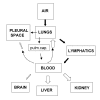
Block diagram to show primary (black arrows) and secondary (white arrows) asbestos fibers translocation. Size of arrows for secondary translocation are representative of accumulation. Accumulation increases in proportion to blood perfusion (lung, kidney and liver) and microvascular permeability (liver), while it is lower in brain and pleural space due to low permeability of the blood-brain barrier and of the endothelial-parietal mesothelium complex, respectively.

References

    1. Suzuki Y, Kohyama N. Translocation of inhaled asbestos fibers from the lung to other tissues. Am J Ind Med. 1991;19:701–704. doi: 10.1002/ajim.4700190603. - DOI - PubMed
    1. Dodson RF, O'Sullivan MF, Huang J, Holiday DB, Hammar SP. Asbestos in extrapulmonary sites* Omentum and Mesentery. Chest. 2000;117:486–493. doi: 10.1378/chest.117.2.486. - DOI - PubMed
    1. Suzuki Y, Yuen SR, Ashley R. Short thin asbestos fibers contribute to the development of human malignant mesothelioma: pathological evidence. Int J Hyg Environ-Health. 2005;208:201–210. doi: 10.1016/j.ijheh.2005.01.015. - DOI - PubMed
    1. Miserocchi G, Venturoli D, Negrini D, Del Fabbro M. Model of pleural fluid turnover. J Appl Physiol. 1993;75:1798–806. - PubMed
    1. Miserocchi G, Negrini D. Pleural space: pressures and fluid dynamics. In: Crystal RG, West JB Weibel ER, Barnes PJ, editor. The Lung: Scientific Foundations. 2. Vol. 1. Philadelphia: Lippincott-Raven Pub; 1997. pp. 1217–1225.

Publication types