. 2008 Jan 24:9:4.
doi: 10.1186/1471-2121-9-4.

The androgen receptor can signal through Wnt/beta-Catenin in prostate cancer cells as an adaptation mechanism to castration levels of androgens

Affiliations

The androgen receptor can signal through Wnt/beta-Catenin in prostate cancer cells as an adaptation mechanism to castration levels of androgens

Liang Schweizer et al. BMC Cell Biol. .

Abstract

Background: A crucial event in Prostate Cancer progression is the conversion from a hormone-sensitive to a hormone-refractory disease state. Correlating with this transition, androgen receptor (AR) amplification and mutations are often observed in patients failing hormonal ablation therapies. beta-Catenin, an essential component of the canonical Wnt signaling pathway, was shown to be a coactivator of the AR signaling in the presence of androgens. However, it is not yet clear what effect the increased levels of the AR could have on the Wnt signaling pathway in these hormone-refractory prostate cells.

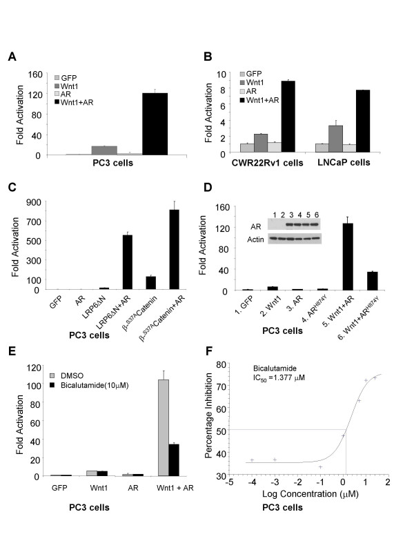
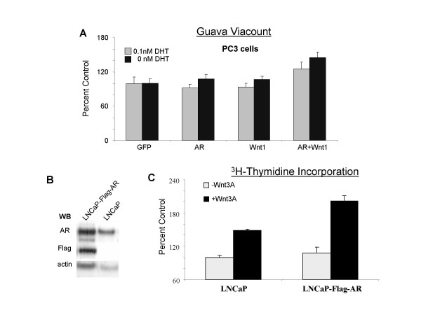
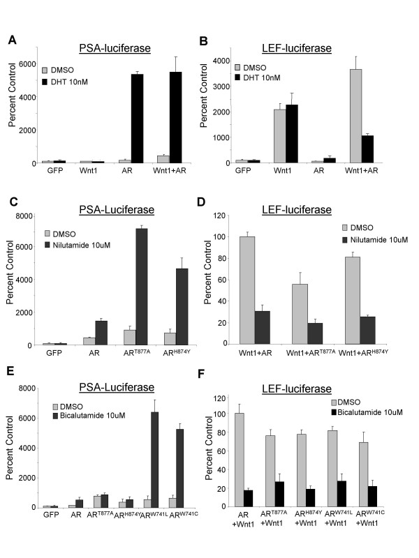
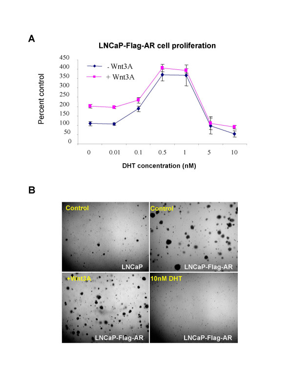
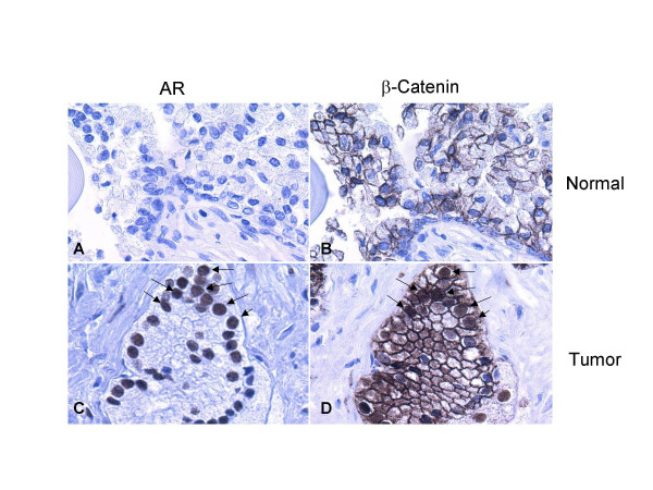
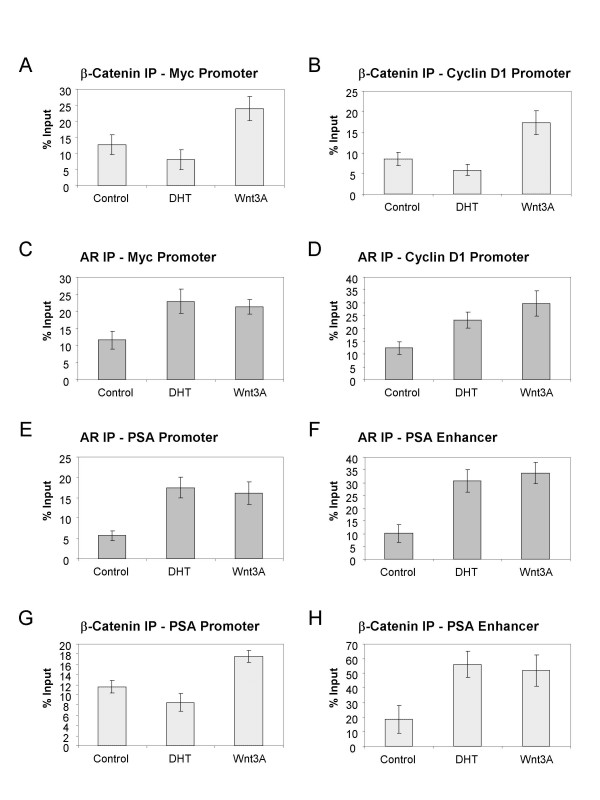
Results: Transient transfections of several human prostate cancer cell lines with the AR and multiple components of the Wnt signaling pathway demonstrate that the AR overexpression can potentiate the transcriptional activities of Wnt/beta-Catenin signaling. In addition, the simultaneous activation of the Wnt signaling pathway and overexpression of the AR promote prostate cancer cell growth and transformation at castration levels of androgens. Interestingly, the presence of physiological levels of androgen or other AR agonists inhibits these effects. These observations are consistent with the nuclear co-localization of the AR and beta-Catenin shown by immunohistochemistry in human prostate cancer samples. Furthermore, chromatin immunoprecipitation assays showed that Wnt3A can recruit the AR to the promoter regions of Myc and Cyclin D1, which are well-characterized downstream targets of the Wnt signalling pathway. The same assays demonstrated that the AR and beta-Catenin can be recruited to the promoter and enhancer regions of a known AR target gene PSA upon Wnt signaling. These results suggest that the AR is promoting Wnt signaling at the chromatin level.

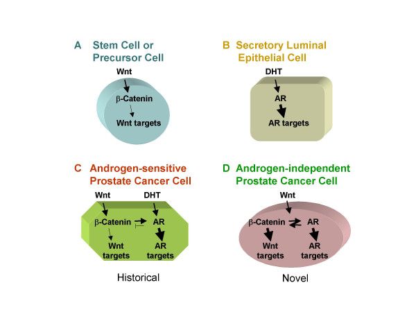
Conclusion: Our findings suggest that the AR signaling through the Wnt/beta-Catenin pathway should be added to the well established functional interactions between both pathways. Moreover, our data show that via this interaction the AR could promote prostate cell malignancy in a ligand-independent manner.


Figures

Figure 1
Figure 1
The overexpression of wild-type or mutant AR potentiates Wnt signaling. Cells grown in RPMI with 10% FBS were transiently transfected with indicated plasmids together with LEF-luciferase and pCMV-Renilla reporter constructs in 12 well plates. Cotransfection of Wnt1 and AR leads to a synergistic effect on the activation of LEF-luciferase reporter in PC3 cells (A), CWR22Rv1 and LNCaP cells (B), compared with transfection with Wnt1 alone. The AR can also potentiate the activation of Wnt signaling in cells expressing a constitutively activated human receptor LRP6, truncated at the N terminus (H6ΔN), or a stabilized β-Catenin S37A (C). The synergistic effect between AR mutants and Wnt1 signaling is shown (D). AR expression levels were measured for both wild type and mutant ARH874Y. Actin levels were used as loading controls. The effect of an AR antagonist is shown (E & F). PC3 cells were treated with bicalutamide 4 hours after transfection. Dual luciferase assay was performed 20–24 hours after treatment.
Figure 2
Figure 2
The synergy of Wnt and AR promotes proliferation in prostate cancer cells. (A) PC3 cells in phenol-red free RPMI with 5% charcoal-stripped serum were transfected with the indicated plasmids and treatments in 96 well plates. 7 days after transfection, the number of cells was determined with a Guava proliferation assay. Cells transfected with GFP were used as control. The percentage of cell numbers was calculated for cells transfected with Wnt or AR or both compared with control samples. (B) AR expression and Flag-tagged AR in LNCaP-Flag-AR cells and LNCaP cells were detected by Western blot. Actin was used as loading control. (C). LNCaP cells and LNCaP-Flag-AR cells in phenol-red free RPMI with 5% charcoal-stripped serum were treated with 100 ng/ml Wnt3A for four days in 96 well plates. The cells were then labelled with [3H]-thymidine and harvested to measure the amount of thymidine incorporation. LNCaP cells which received no Wnt3A treatment were used as control. The percentage of [3H]-thymidine incorporation was calculated compared with control.
Figure 3
Figure 3
AR agonists inhibit the Wnt and AR synergy. PC3 cells in phenol-red free RPMI with 5% charcoal-stripped serum in 96 well plates were transfected with the indicated plasmids, together with pCMV-Renilla reporter and PSA-luciferase (A, C, & E) or LEF-Luciferase (B, D, & F). The following treatments were applied to cells 4 hours after transfection: 10 nM DHT (A & B); 10 uM Nilutamide (C & D); 10 uM Bicalutamide (E & F). Dual luciferase assays were performed 20–24 hours after treatment.
Figure 4
Figure 4
The effect of DHT on cell proliferation and soft agar growth. (A) LNCaP-Flag-AR cells in phenol-red free RPMI with 5% charcoal-stripped serum were treated with DHT ranging from 0.01 nM to 10 nM with or without Wnt3A 100 ng/ml for four days in 96 well plates. The cells were then labeled with [3H]-thymidine and harvested to measure the thymidine incorporation. Cells that received no DHT treatment were used as control. The percentage of [3H]-thymidine incorporation was calculated compared with control. (B). LNCaP and LNCaP-Flag-AR cells were plated in soft agar with no treatment as control or with 100 ng/ml Wnt3A, or 10 nM DHT. After approximately 4 weeks, colonies were fixed with 10% formaldehyde in PBS. A representative field of cells was photographed for each cell type, with or without treatment using bright-field microscopy. Upper panel LNCaP or LNCaP-Flag-AR cells received no treatment, while lower panel, LNCaP-Flag-AR cells were either treated with Wnt3A or DHT.
Figure 5
Figure 5
Expression of AR and β-Catenin in normal prostate tissues and tumor samples. A tissue microarray containing 9 prostate tumors and 4 normal prostate tissue samples was stained with antibodies against AR and β-Catenin. In normal prostate tissues, the low levels of AR expression, compared with the high levels in prostate cancer cells, were indicated by lack of AR staining in cells (A); β-Catenin was predominantly located at the normal cytoplasmic membrane (B). In late stage prostate cancer samples, AR was substantially overexpressed in the nuclei of the prostate cancer cells (C) where nuclear β-Catenin staining was also observed in some of these cells (D).
Figure 6
Figure 6
The recruitment of AR and β-Catenin to Wnt signaling target genes as well as PSA promoter and enhancer region using CHIP analysis. LNCaP cells in phenol-red free RPMI with 5% charcoal-stripped serum were treated with either 10 nM DHT, 100 ng/ml Wnt3A, or no treatment for 16 hours before cross-linking. Anti-β-Catenin (A, B, G and H) and anti-AR (C, D, E and F) antibodies, together with negative control IgG were then used for immunoprecipitation. After reverse crosslinking and DNA purification, PCR products were analyzed using the Agilent 2100 bioanalyzer. Percent of input is shown here to compare the levels of β-Catenin or AR at the promoter or enhancer region of PSA, or the promoter region of Cyclin D1 or Myc.
Figure 7
Figure 7
Working model for the interaction of AR and Wnt/β-Catenin signaling pathway in castration-resistant prostate cancer cells. In normal conditions, Wnt and androgen signaling function at different cell populations (A&B). In prostate cancer where both pathways may be deregulated in the same cells, androgens may inhibit the input of AR into the Wnt signaling pathway where β-Catenin can potentiate the AR signaling (C). In castration-resistant prostate cancer cells, Wnt can activate both signaling pathways turning the unliganded AR into a coactivator of the Wnt dependent transcriptional program (D).

References

    1. Koivisto P, et al. Androgen receptor gene amplification: a possible molecular mechanism for androgen deprivation therapy failure in prostate cancer. Cancer Res. 1997;57:314–9. - PubMed
    1. Linja MJ, et al. Amplification and Overexpression of Androgen Receptor Gene in Hormone-Refractory Prostate Cancer. Cancer Res. 2001;61:3550–3555. - PubMed
    1. Chen CD, et al. Molecular determinants of resistance to antiandrogen therapy. Nat Med. 2004;10:33–39. doi: 10.1038/nm972. - DOI - PubMed
    1. Grossmann ME, Huang H, Tindall DJ. Androgen receptor signaling in androgen-refractory prostate cancer. J Natl Cancer Inst. 2001;93:1687–97. doi: 10.1093/jnci/93.22.1687. - DOI - PubMed
    1. Buchanan G, et al. Mutations at the boundary of the hinge and ligand binding domain of the androgen receptor confer increased transactivation function. Mol Endocrinol. 2001;15:46–56. doi: 10.1210/me.15.1.46. - DOI - PubMed

MeSH terms