

Constitutive overexpression of a novel 21 kDa protein by Hodgkin lymphoma and aggressive non-Hodgkin lymphomas
- PMID: 18218123
- PMCID: PMC2267462
- DOI: 10.1186/1476-4598-7-12
Constitutive overexpression of a novel 21 kDa protein by Hodgkin lymphoma and aggressive non-Hodgkin lymphomas
Abstract
Background: CD30, a 120 kDa surface phosphorylated protein is a member of tumour necrosis/nerve growth factor receptor (TNF/NGFR) family and constitutively expressed by Hodgkin and Reed-Sternberg (HRS) cells of Hodgkin lymphoma (HL) and the neoplastic cells of Anaplastic Large Cell Lymphoma (ALCL). A disease-specific protein marker is yet to be identified in Hodgkin lymphoma cells. In order to define HL-specific biomarkers, novel murine monoclonal antibodies were developed in our laboratory.
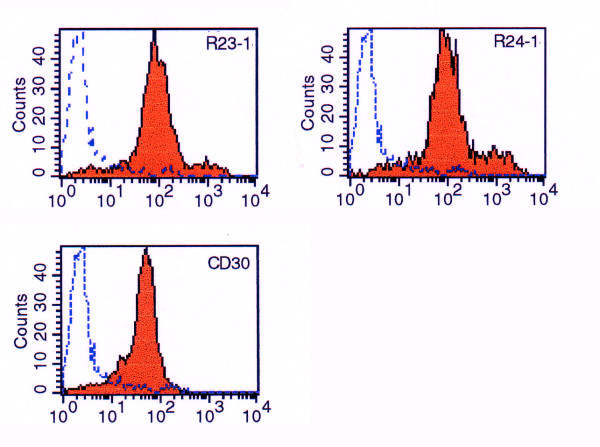
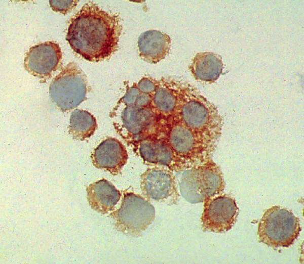
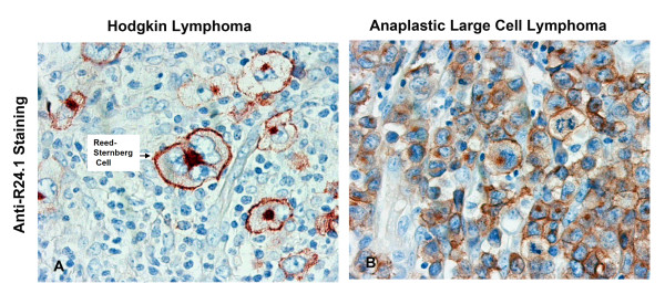
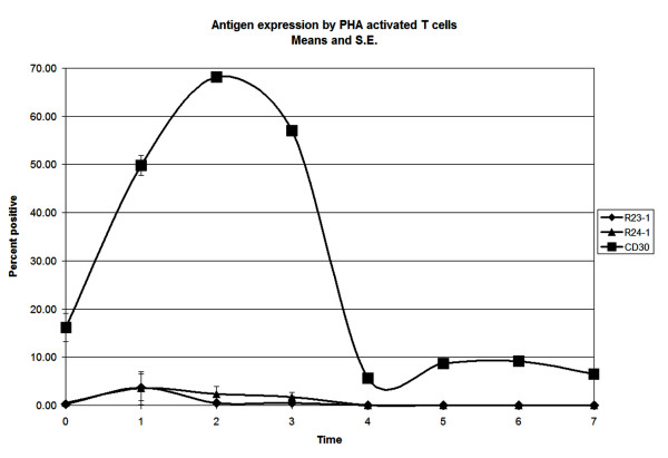
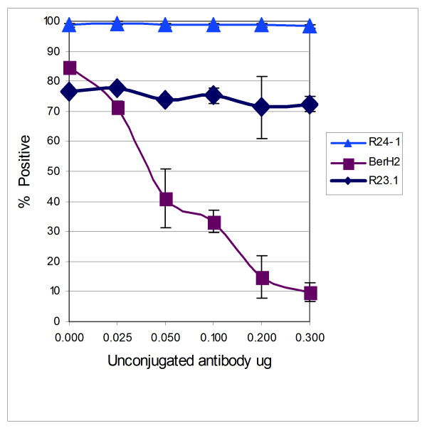
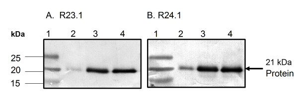
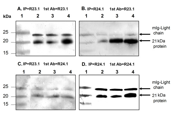
Results: Murine monoclonal antibodies (mabs) were raised against the B3 sub clone of HL-derived cell line KM-H2. Two of these mabs (clone R23.1 mab and clone R24.1 mab) are IgG1 class antibodies that recognize a 21 kDa protein present at the cell membrane and in the cytoplasm in HL-derived cell lines. Clone R24.1 mab recognizes a formalin-resistant epitope and labels HRS cells in tissue samples from patients with HL of the classical type, ALCL, and subsets of T and B cell aggressive Non-Hodgkin Lymphomas (NHL). The antigen recognized by the clone R23.1 mab and clone R24.1 mab does not share epitopes with CD30 cluster regions A, B, or C, and, unlike CD30, is not expressed by phytohemagglutinin (PHA) activated T cells.
Conclusion: The 21 kDa protein detected by clone R23.1 and clone R24.1 mabs is a novel membrane-associated protein that may be a potential marker for the diagnosis and targeted therapy of HL and aggressive T and B cell NHL.
Figures
Similar articles
-
Constitutively overexpressed 21 kDa protein in Hodgkin lymphoma and aggressive non-Hodgkin lymphomas identified as cytochrome B5b (CYB5B).Mol Cancer. 2010 Jan 26;9:14. doi: 10.1186/1476-4598-9-14. Mol Cancer. 2010. PMID: 20100355 Free PMC article.
-
Signal transducer and activator of transcription 6 is frequently activated in Hodgkin and Reed-Sternberg cells of Hodgkin lymphoma.Blood. 2002 Jan 15;99(2):618-26. doi: 10.1182/blood.v99.2.618. Blood. 2002. PMID: 11781246
-
CD30 ligand, a member of the TNF ligand superfamily, with growth and activation control CD30+ lymphoid and lymphoma cells.Leuk Lymphoma. 1996 Feb;20(5-6):397-409. doi: 10.3109/10428199609052421. Leuk Lymphoma. 1996. PMID: 8833395 Review.
-
Expression of cytotoxic proteins in peripheral T-cell and natural killer-cell (NK) lymphomas: association with extranodal site, NK or Tgammadelta phenotype, anaplastic morphology and CD30 expression.Leuk Lymphoma. 2000 Jul;38(3-4):317-26. doi: 10.3109/10428190009087022. Leuk Lymphoma. 2000. PMID: 10830738 Review.
-
The Role of Reed-Sternberg CD30 Receptor and Lymphocytes in Pathogenesis of Disease and Its Implication for Treatment.Acta Med Indones. 2018 Apr;50(2):93-95. Acta Med Indones. 2018. PMID: 29950526
Cited by
-
Proteomic analysis of serum in lung cancer induced by 3-methylcholanthrene.J Biomed Biotechnol. 2009;2009:397910. doi: 10.1155/2009/397910. Epub 2009 Sep 24. J Biomed Biotechnol. 2009. PMID: 19794824 Free PMC article.
-
Constitutively overexpressed 21 kDa protein in Hodgkin lymphoma and aggressive non-Hodgkin lymphomas identified as cytochrome B5b (CYB5B).Mol Cancer. 2010 Jan 26;9:14. doi: 10.1186/1476-4598-9-14. Mol Cancer. 2010. PMID: 20100355 Free PMC article.
-
Recent Advances in the Pathobiology of Hodgkin's Lymphoma: Potential Impact on Diagnostic, Predictive, and Therapeutic Strategies.Adv Hematol. 2011;2011:439456. doi: 10.1155/2011/439456. Epub 2011 Jan 18. Adv Hematol. 2011. PMID: 21318045 Free PMC article.
-
Nuclear Factor kappa B is central to Marek's disease herpesvirus induced neoplastic transformation of CD30 expressing lymphocytes in-vivo.BMC Syst Biol. 2012 Sep 14;6:123. doi: 10.1186/1752-0509-6-123. BMC Syst Biol. 2012. PMID: 22979947 Free PMC article.
References
-
- Schwarting R, Gerdes J, Durkop H, Falini B, Pileri S, Stein H. BER-H2: a new anti-Ki-1 (CD30) monoclonal antibody directed at a formol-resistant epitope. Blood. 1989;74:1678–1689. - PubMed
-
- Horn-Lohrens O, Tiemann M, Lange H, Kobarg J, Hafner M, Hansen H, Sterry W, Parwaresch RM, Lemke H. Shedding of the soluble form of CD30 from the Hodgkin-analogous cell line L540 is strongly inhibited by a new CD30-specific antibody (Ki-4) Int J Cancer. 1995;60:539–544. doi: 10.1002/ijc.2910600419. - DOI - PubMed
-
- Stein H, Mason DY, Gerdes J, O'Connor N, Wainscoat J, Pallesen G, Gatter K, Falini B, Delsol G, Lemke H, Schwarting R, Lennert k. The expression of the Hodgkin's disease associated antigen Ki-1 in reactive and neoplastic lymphoid tissue: evidence that Reed-Sternberg cells and histiocytic malignancies are derived from activated lymphoid cells. Blood. 1985;66:848–858. - PubMed
-
- Delsol G, Al Saati T, Gatter KC, Gerdes J, Schwarting R, Caveriviere P, Rigal-Huguet F, Robert A, Stein H, Mason DY. Coexpression of epithelial membrane antigen (EMA), Ki-1, and interleukin-2 receptor by anaplastic large cell lymphomas. Diagnostic value in so-called malignant histiocytosis. Am J Pathol. 1988;130:59–70. - PMC - PubMed
MeSH terms
Substances
LinkOut - more resources
Full Text Sources
Other Literature Sources
Medical
