. 2008 Feb;14(2):45-53.
doi: 10.1016/j.molmed.2007.12.002. Epub 2008 Jan 22.

Amyloid beta, mitochondrial dysfunction and synaptic damage: implications for cognitive decline in aging and Alzheimer's disease

Affiliations

Amyloid beta, mitochondrial dysfunction and synaptic damage: implications for cognitive decline in aging and Alzheimer's disease

P Hemachandra Reddy et al. Trends Mol Med. 2008 Feb.

Abstract

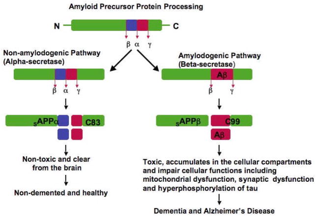
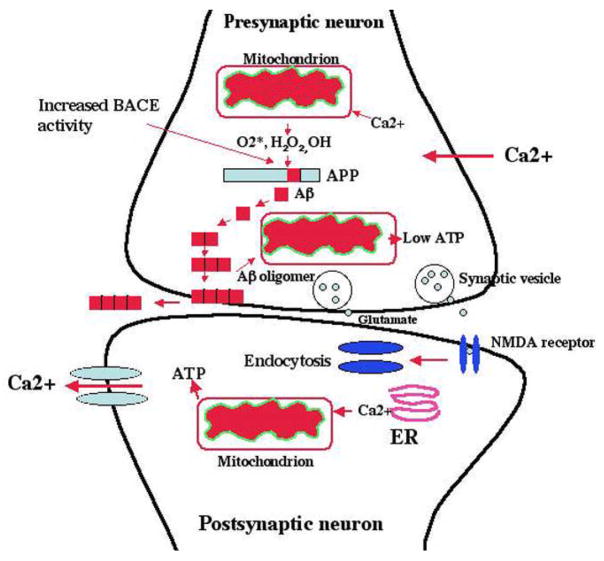
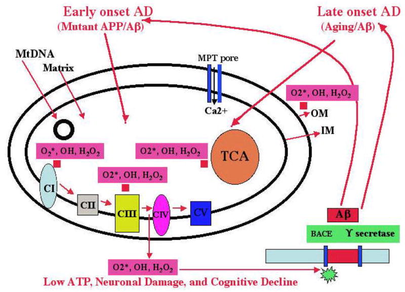
Recent studies of postmortem brains from Alzheimer's disease (AD) patients and transgenic mouse models of AD suggest that oxidative damage, induced by amyloid beta (Abeta), is associated with mitochondria early in AD progression. Abeta and amyloid-precursor protein are known to localize to mitochondrial membranes, block the transport of nuclear-encoded mitochondrial proteins to mitochondria, interact with mitochondrial proteins, disrupt the electron-transport chain, increase reactive oxygen species production, cause mitochondrial damage and prevent neurons from functioning normally. Furthermore, accumulation of Abeta at synaptic terminals might contribute to synaptic damage and cognitive decline in patients with AD. Here, we describe recent studies regarding the roles of Abeta and mitochondrial function in AD progression and particularly in synaptic damage and cognitive decline.


Figures

Figure 1
Figure 1. APP processing in non-demented healthy individuals and AD patients
APP processing occurs via 2 pathways. Beta secretase based amyloidogenic and α-secretase based non-amyloidogenic: In non-amyloidogenic pathway, cleavage occurs by α-secretase within the Aβ domain and generates the large soluble N-terminal fragment (sAPPα) and a non-amyloidogenic C-terminal fragment of 83 aminoacid residues (C83). Further cleavage of this C-terminal fragment by γ-secretase generates the non-amyloidogenic peptide (P3) and APP intracellular domain (ACID). These products are nontoxic. The non-amyloidogenic α-secretase pathway occurs in over 90% of humans, and these individuals generally do not develop dementia. In amyloidogenic pathway, cleavage occurs by β-secretase at the beginning of the Aβ domain and generates a soluble N-terminus fragment (sAPPβ , and amyloidogenic C-terminal fragment of 99 residues (C99). This C-terminal fragment, further cleaved by γ-secretase and generates Aβ. Cleavage by multiple γ-secretases can generate Aβ1-40 and Aβ1-42 fragments. Amyloid beta can accumulate in cellular compartments such as mitochondria and lysosomes and impair cellular functions. Amyloidogenic pathway occurs in about 10% of total humans and these individuals may develop dementia and AD. Age-dependent decrease of Aβ degrading enzymes such as neprilysin and insulin degrading enzyme may not clear Aβ in neurons that contribute to Aβ accumulation in the brain.
Figure 2
Figure 2. Hypothesized changes at the synaptic mitochondria in AD neurons
In AD neurons, Aβ (1–40 and 1–42) are secreted via sequential cleavage of Aβ precursor protein (APP) by beta secretase (BACE) and gamma secretase. In late-onset AD, mitochondrially produced free radicals (O2*, H2O2, OH) activate beta secretase and facilitate the cleavage of APP. The cleaved Aβ may further enter mitochondria primarily localized at synapses, induce free radicals, cause oxidative damage, and inhibit cellular ATP. Increased levels of BACE activity have been observed in the postmortem brain specimens from late-onset AD patients. This increased BACE activity may be primarily due to decreased energy metabolism – in other words, it may be due to decreased ATP, increased Ca2+ influx, and increased free radicals. Reduced cellular ATP production, in turn, may cause the impairment of neurotransmission (glutamate and NMDA receptors). These events may occur primarily in learning and memory regions of the brain, and ultimately may cause cognitive decline in AD patients.
Figure 3
Figure 3. Mitochondrial hypothesis and AD
In early-onset AD, mutant APP and soluble Aβ are hypothesized to localize to synaptic mitochondria, leading to the generation of free radicals (O2*, H2O2, OH). The free radicals, in turn, may decrease cytochrome oxidase activity and inhibit cellular ATP. In late-onset AD, the free radicals that are generated due to aging may activate BACE and facilitate the cleavage of the Aβ. The cleaved Aβ may enter mitochondria and induce free radicals (O2*, H2O2, OH), leading to the disruption of the ETC, a decrease in cytochrome oxidase activity, and the inhibition of ATP. This feedback loop may ultimately lead to neuronal damage, to the degeneration of neurons, and to cognitive decline in AD patients.

Similar articles

Cited by

References

    1. Manczak M, et al. Mitochondria are a direct site of Abeta accumulation in Alzheimer's disease neurons: implications for free radical generation and oxidative damage in disease progression. Hum Mol Genet. 2006;15:1437–1449. - PubMed
    1. Devi L, et al. Accumulation of amyloid precursor protein in the mitochondrial import channels of human Alzheimer's disease brain is associated with mitochondrial dysfunction. J Neurosci. 2006;26:9057–9068. - PMC - PubMed
    1. Caspersen C, et al. Mitochondrial Abeta: a potential focal point for neuronal metabolic dysfunction in Alzheimer's disease. FASEB J. 2005;19:2040–1. - PubMed
    1. Nunomura A, et al. Oxidative damage is the earliest event in Alzheimer disease. J Neuropathol Exp Neurol. 2001;60:759–767. - PubMed
    1. Selkoe DJ. Alzheimer's disease is a synaptic failure. Science. 2002;298:789–791. - PubMed

Publication types

Substances