. 2008 Mar 28;283(13):8624-33.
doi: 10.1074/jbc.M707860200. Epub 2008 Jan 23.

Anti-cancer effects of JKA97 are associated with its induction of cell apoptosis via a Bax-dependent and p53-independent pathway

Affiliations

Anti-cancer effects of JKA97 are associated with its induction of cell apoptosis via a Bax-dependent and p53-independent pathway

Wenjing Luo et al. J Biol Chem. .

Erratum in

  • J Biol Chem. 2012 Jul 6;287(28):23850

Abstract

p53, one of the most commonly mutated genes in human cancers, is thought to be associated with cancer development. Hence, screening and identifying natural or synthetic compounds with anti-cancer activity via p53-independent pathway is one of the most challenging tasks for scientists in this field. Compound JKA97 (methoxy-1-styryl-9H-pyrid-[3,4-b]-indole) is a small molecule synthetic anti-cancer agent, with unknown mechanism(s). In this study we have demonstrated that the anti-cancer activity of JKA97 is associated with apoptotic induction via p53-independent mechanisms. We found that co-incubation of human colon cancer HCT116 cells with JKA97 inhibited HCT116 cell anchorage-independent growth in vitro and tumorigenicity in nude mice and also induced a cell apoptotic response, both in the cell culture model and in a tumorigenesis nude mouse model. Further studies showed that JKA97-induced apoptosis was dramatically impaired in Bax knock-out (Bax(-/-)) HCT116 cells, whereas the knock-out of p53 or PUMA did not show any inhibitory effects. The p53-independent apoptotic induction by JKA97 was confirmed in other colon cancer and hepatocarcinoma cell lines. In addition, our results showed an induction of Bax translocation and cytochrome c release from the mitochondria to the cytosol in HCT116 cells, demonstrating that the compound induces apoptosis through a Bax-initiated mitochondria-dependent pathway. These studies provide a molecular basis for the therapeutic application of JKA97 against human cancers with p53 mutations.


Figures

FIGURE 1.
FIGURE 1.
The structure of JKA97.
FIGURE 2.
FIGURE 2.
JKA97 inhibits HCT116 cell anchorage-independent growth. HCT116 cells (1 × 10⁴) were exposed to JKA97 (0-10 μm) in 0.33% agar for 14 days as described under “Materials and Methods.” The number of colonies was counted under microscopy at the end of the experiments (A). The colonies are expressed as means ± S.E. from six assays. *, p < 0.05, significant increase from vehicle control (B).
FIGURE 3.
FIGURE 3.
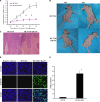
JKA97 Inhibits HCT116 cell tumorigenicity in nude mice. A and B, nude mice 6 weeks of age were used. The experiments were conducted with 16 mice in each group. HCT116 cells were injected subcutaneously into the flanks of each mouse to initiate tumor growth for 14 days. The mice were then treated intraperitoneally with either control vehicle or JKA97 once a day, 5 days a week, for a total of 5 weeks as indicated under “Materials and Methods.” Tumor size was measured weekly in two dimensions throughout the study. Data are shown as the means ± S.E. (A) and external appearance of tumors (B). *, p < 0.05, significant increase from vehicle control. Tumors were removed from mice at week 7 after HCT116 cell injection, fixed in 10% buffered formalin, and embedded in paraffin. C, 4-μm sections were dehydrated and stained with hematoxylin and eosin. The slides were observed under microscope and photographed (original magnification, ×400). D, paraffin-embedded tumors xenografts were sectioned (4 μm) and subjected to TUNEL staining as described under “Materials and Methods.” Three slides from each tumor sample were used for TUNNEL assay. The slides were then observed under a fluorescent microscope and photographed (original magnification, ×400). The apoptotic cells (green) were stained by the TUNEL assay. Cell nuclei (blue) were stained with Hoechst. E, average percentage of TUNEL positive cells in tumors. Data are shown as the means ± S.E. *, p < 0.05, significant increase from vehicle control group.
FIGURE 4.
FIGURE 4.
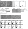
JKA97 induces cell death in human colon cancer HCT116 and human hepatocarcinoma cell line SMMC-7721. A, HCT116 cells (4 × 10⁵) were exposed to JKA97 (0 or 10 μm) for 24 h, and the morphological changes were observed under microscope and photographed (original magnification, ×400). B, HCT116 cells (4× 10⁵) were seeded into each well of 6-well plates and cultured until the cell density reached 70-80% confluence. The cells were then exposed to 0 or 10 μm JKA97 for 24 h. The cell death was determined using PI staining and detected by flow cytometry. C, cleavage of caspase 3 and PARP was detected by Western blotting (12 h) after HCT116 cells were exposed to various doses of JKA97. β-Actin was used as the protein loading control. D, SMMC-7721 cells (4× 10⁵) were exposed to JKA97 (0 or 10 μm) for 24 h, and the morphological changes were observed under an inverting microscope and photographed (original magnification, ×400). E, the cleavage of caspase 3 was detected in JKA97-treated SMMC-7721 cells by Western blotting at 12 h after JKA97 exposure. β-Actin was used as the protein loading control. F, the HCT116 cells were pretreated with Z-VAD-fmk (25 mm) and caspase 3 inhibitor VII (20 nm) for 30 min and then exposed to 10 μm JKA97 for 24 h; the morphological changes were observed under a microscope and photographed (original magnification, ×400). G, JKA97-induced cell death was determined using Annexin-V staining and detected by flow cytometry.
FIGURE 5.
FIGURE 5.
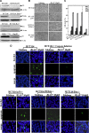
JKA97-induced HCT116 cell apoptosis was through Bax-dependent and p53- and PUMA-independent mechanisms. A, HCT116 (wild type), HCT116-p53-/-, HCT116-PUMA-/-, HCT116-Bax-/-, SMMC7721, and Hep3B cells were analyzed for phenotype identification by Western blotting with specific antibodies as indicated. β-Actin was used as the protein loading control. B, HCT116, HCT116-p53-/-, HCT116-PUMA-/-, and HCT116-Bax-/- cells were plated in 96-well plates (1 × 10⁴ cells/well) in McCoy's 5A medium supplemented with 10% fetal bovine serum. 24 h later, the medium was replaced with 0.1% serum, and the cells were exposed to either vehicle control or 10 μm JKA97 for 24 h. The morphological changes were observed under microscope and photographed (original magnification, ×400). C-E, HCT116, HCT116-p53-/-, HCT116-PUMA-/-, and HCT116-Bax-/- cells were exposed to vehicle control or 10 μm JKA97 for 24 h. Cell death was determined by MTS assay (C), TUNEL assay (D), and a PI-staining flow cytometry assay (E). F, caspase 3 and PARP activation and cleavage were detected by Western blotting (12 h). β-Actin was used as the protein loading control. G, SMMC-7721 and Hep3B cells were plated in 6-well plates and cultured until the cell density reached 70-80% confluence. The cells were exposed to vehicle control or 10 μm JKA97 in 0.1% FBS medium for 24 h. The morphological changes were observed under a microscope and photographed (original magnification, ×400). H, JKA97-induced apoptosis was detected by determination of cleavage of caspase 3 at 12 h after SMMC-7721 and Hep3B cells were exposed to 10 μm JKA97. I and J, JKA97-treated cell apoptosis was determined with Annexin-V staining (I) and a TUNEL assay (J) in SMMC-7721, Hep3B, and SW620 cells.
FIGURE 5.
FIGURE 5.
JKA97-induced HCT116 cell apoptosis was through Bax-dependent and p53- and PUMA-independent mechanisms. A, HCT116 (wild type), HCT116-p53-/-, HCT116-PUMA-/-, HCT116-Bax-/-, SMMC7721, and Hep3B cells were analyzed for phenotype identification by Western blotting with specific antibodies as indicated. β-Actin was used as the protein loading control. B, HCT116, HCT116-p53-/-, HCT116-PUMA-/-, and HCT116-Bax-/- cells were plated in 96-well plates (1 × 10⁴ cells/well) in McCoy's 5A medium supplemented with 10% fetal bovine serum. 24 h later, the medium was replaced with 0.1% serum, and the cells were exposed to either vehicle control or 10 μm JKA97 for 24 h. The morphological changes were observed under microscope and photographed (original magnification, ×400). C-E, HCT116, HCT116-p53-/-, HCT116-PUMA-/-, and HCT116-Bax-/- cells were exposed to vehicle control or 10 μm JKA97 for 24 h. Cell death was determined by MTS assay (C), TUNEL assay (D), and a PI-staining flow cytometry assay (E). F, caspase 3 and PARP activation and cleavage were detected by Western blotting (12 h). β-Actin was used as the protein loading control. G, SMMC-7721 and Hep3B cells were plated in 6-well plates and cultured until the cell density reached 70-80% confluence. The cells were exposed to vehicle control or 10 μm JKA97 in 0.1% FBS medium for 24 h. The morphological changes were observed under a microscope and photographed (original magnification, ×400). H, JKA97-induced apoptosis was detected by determination of cleavage of caspase 3 at 12 h after SMMC-7721 and Hep3B cells were exposed to 10 μm JKA97. I and J, JKA97-treated cell apoptosis was determined with Annexin-V staining (I) and a TUNEL assay (J) in SMMC-7721, Hep3B, and SW620 cells.
FIGURE 6.
FIGURE 6.
Determination of protein translocation between cytosol and mitochondria in HCT116 cells exposed to JKA97. A, HCT116 cells grown in 150-mm culture dishes were exposed to vehicle control or 10 μm JKA97. The cytosolic and mitochondrial proteins were isolated, respectively, according to the protocol provided by the mitochondria isolation kit. Equal amounts of cytosolic and mitochondrial proteins isolated from HCT116 cells were separated on 12% SDS-polyacrylamide gels and then subjected to Western blotting analysis for various antibodies as indicated. β-Actin was used as the protein loading control. B, HCT116 cells were exposed to vehicle control or 10 μm JKA97 for 12 or 24 h, and mitochondrial protein were isolated. Bax, Bak, and AIF (apoptosis-inducing factor) were detected by Western blotting. β-Actin was used as the protein loading control.

Similar articles

Cited by

References

    1. Svec, J. (2005) Bratisl. Lek. Listy 106 238-239 - PubMed
    1. Lane, D. P. (1993) Nature 362 786-787 - PubMed
    1. Wang, J., Ouyang, W., Li, J., Wei, L., Ma, Q., Zhang, Z., Tong, Q., He, J., and Huang, C. (2005) Cancer Res. 65 6601-6611 - PubMed
    1. Tweddle, D. A., Pearson, A. D., Haber, M., Norris, M. D., Xue, C., Flemming, C., and Lunec, J. (2003) Cancer Lett. 197 93-98 - PubMed
    1. Cheng, J., and Haas, M. (1990) Mol. Cell. Biol. 10 5502-5509 - PMC - PubMed

Publication types

MeSH terms