. 2008 Mar 28;283(13):8756-64.
doi: 10.1074/jbc.M708223200. Epub 2008 Jan 24.

Kv1.5 association modifies Kv1.3 traffic and membrane localization

Affiliations

Kv1.5 association modifies Kv1.3 traffic and membrane localization

Rubén Vicente et al. J Biol Chem. .

Abstract

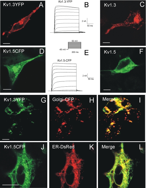
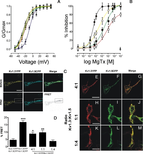
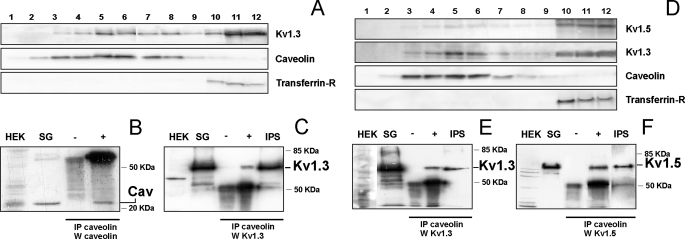
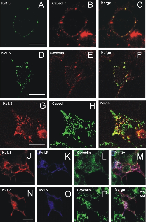
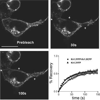
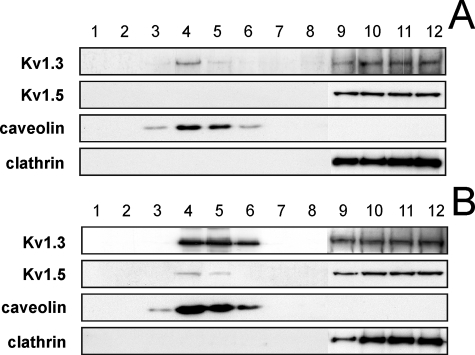
Kv1.3 activity is determined by raft association. In addition to Kv1.3, leukocytes also express Kv1.5, and both channels control physiological responses. Because the oligomeric composition may modify the channel targeting to the membrane, we investigated heterotetrameric Kv1.3/Kv1.5 channel traffic and targeting in HEK cells. Kv1.3 and Kv1.5 generate multiple heterotetramers with differential surface expression according to the subunit composition. FRET analysis and pharmacology confirm the presence of functional hybrid channels. Raft association was evaluated by cholesterol depletion, caveolae colocalization, and lateral diffusion at the cell surface. Immunoprecipitation showed that both Kv1.3 and heteromeric channels associate with caveolar raft domains. However, homomeric Kv1.3 channels showed higher association with caveolin traffic. Moreover, FRAP analysis revealed higher mobility for hybrid Kv1.3/Kv1.5 than Kv1.3 homotetramers, suggesting that heteromers target to distinct surface microdomains. Studies with lipopolysaccharide-activated macrophages further supported that different physiological mechanisms govern Kv1.3 and Kv1.5 targeting to rafts. Our results implicate the traffic and localization of Kv1.3/Kv1.5 heteromers in the complex regulation of immune system cells.


Figures

FIGURE 1.
FIGURE 1.
Kv1.3-YFP and Kv1.5-CFP have distinct cellular distributions. HEK cells were transient transfected with either wild type Kv1.3 (C) and Kv1.5 (F) or Kv1.3-YFP (A and B) and Kv1.5-CFP (D and E). Representative traces of K+ currents from Kv1.3-YFP (B) and Kv1.5-CFP cells (E) are shown. The cells were held at -60 mV, and currents were elicited by depolarizing pulses in 10-mV steps (200 ms in duration) from -50 to +50 mV. C, immunolocalization of untagged Kv1.3. F, untagged Kv1.5. G-I, Kv1.3-YFP cotransfected with Golgi-CFP marker. J-L, Kv1.5-CFP cotransfected with DsRed-ER marker. I and L, merge panels show colocalization (yellow). The bars represent 10 μm.
FIGURE 2.
FIGURE 2.
Kv1.3 associates with Kv1.5, leading to biophysically and pharmacologically distinct channels. HEK cells were doubly transfected with different ratios of Kv1.3-YFP and Kv1.5-CFP. A, plot of normalized conductance versus voltage of K+ currents from HEK cells expressing different channel ratios. The pulse protocols are shown in Fig. 1. The conductance was normalized to the peak current at +50 mV. B, inhibition of the K+ current by Margatoxin. The currents were evoked at +50 mV from a holding potential of -60 mV during a pulse potential of 200 ms. The percentage of inhibition was calculated by comparing the current at a given concentration of toxin versus that obtained in its absence. The values are the means ± S.E. The symbols and color lines are: Kv1.3-YFP (black circles, black line), Kv1.5-CFP (closed triangle, blue line), and the different Kv1.3-YFP/Kv1.5-CFP ratios 4:1 (white circles, yellow line), 1:1 (black squares, green line), and 1:4 (white squares, red line). C, representative FRET experiment. Panels show fluorescence signal of Kv1.3-YFP and Kv1.5-CFP (ratio 1:1) before and after photobleaching of the YFP region bounded by the rectangle. The FRET panel represents the difference of CFP fluorescence intensity after and before photobleaching. D, histogram shows FRET efficiency of different ratios of Kv1.3-YFP and Kv1.5-CFP. Negative control was performed with cells expressing Kv1.5-CFP and Kv2.1-YFP. Positive control was performed using cells expressing Kv1.3-CFP and Kv1.3-YFP. *, p < 0.05; **, p < 0.01; ***, p < 0.001 versus Kv2.1/Kv1.5 (Student's t test). E-M, HEK cells were doubly transfected with different ratios of Kv1.3-YFP (red) and Kv1.5-CFP (green). The merge panels show colocalization (yellow). Kv1.3 and Kv1.5 colocalized in HEK cells with 4:1 (E-G), 1:1 (H-J), and 1:4 (K-M) ratios. The bars represent 10 μm.
FIGURE 3.
FIGURE 3.
Kv1.3 and Kv1.3/Kv1.5 heterotetramers target to lipid rafts. Detergent-based isolation of lipid rafts. Sucrose density gradient centrifugation of 1% Triton X-100-solubilized extracts from cells, transfected with Kv1.3 alone (A-C) or doubly transfected with Kv1.3 and Kv1.5 (D-F), were analyzed by Western blot. While caveolin indicates low buoyancy rafts, transferrin receptor (Transferrin-R) distributes in nonfloating fractions. B and C, fractions 5 and 6 from A were immunoprecipitated (IP) with anti-caveolin antibody, and blots were immunoblotted (W) against caveolin (B) and Kv1.3 (C). E and F, fractions 5 and 6 from D were immunoprecipitated (IP) with anti-caveolin antibody, and blots were immunoblotted (W) against Kv1.3 (E) and Kv1.5 (F). HEK, membrane extracts from nontransfected HEK cells; SG, sucrose fractions number 5 and 6 as starting material; IPS, immunoprecipitated supernatant; -, immunoprecipitation in the absence of anti-caveolin antibody; +, immunoprecipitation in the presence of anti-caveolin antibody.
FIGURE 4.
FIGURE 4.
Cholesterol depletion impairs Kv1.3, Kv1.5, and Kv1.3/Kv1.5 localization in lipid rafts. HEK cells were treated with or without 2% MβCD 1 h before lipid raft extraction (A-D) and confocal microscopy studies (E). A and C, cells were transfected with Kv1.3, Kv1.5, and Kv1.3/Kv1.5 (ratio 1:1) and fractions immunoblotted (WB) against the channels. B and D, quantification of data from A and C. Relative abundance (%) of the protein expression located in floating (white bar) and nonfloating (black bar, Kv1.3; gray bar, Kv1.5) fractions. 100% represents the overall protein expression in all fractions. The values are the means ± S.E. of four independent experiments. *, p < 0.05; **, p < 0.01 (Student's t test). A and B, cells incubated without MβCD (-MβCD). C and D, cells incubated with MβCD (+MβCD). E, MβCD did not alter Kv1.3, Kv1.5, and Kv1.3/Kv1.5 expression patterns. The cells were transfected as described above and incubated in the absence (top panels, -MβCD) or the presence (bottom panels, +MβCD) of MβCD. HEK cells were transfected with Kv1.3-YFP (red), Kv1.5-CFP (green), and both (Kv1.3/Kv1.5). The merge panels show colocalization (yellow) in doubly transfected cells. The bars represent 10 μm.
FIGURE 5.
FIGURE 5.
Kv1.3 and Kv1.5 colocalize with caveolae in HEK cells. A and D, cells expressing Kv1.3 and Kv1.5 patched with their respective antibody against external epitope. B and E, caveolin. C and F, merge panels showed colocalization (yellow). G-I, intracellular Kv1.3 traffics with caveolin. G, Kv1.3-YFP; H, caveolin; I, colocalization (yellow). Hybrid Kv1.3/Kv1.5 channels (ratio 4:1, J-M) but not ratio 1:4 (N-Q) colocalized with intracellular caveolin. J and N, Kv1.3-YFP. K and O, Kv1.5-CFP. L and P, Caveolin. M and Q, colocalization between Kv1.3 and Kv1.5 (pink) or triple colocalization with caveolin (white). The bars represent 10 μm.
FIGURE 6.
FIGURE 6.
FRAP of Kv1.3 and heterotetramer Kv1.3/Kv1.5. FRAP experiments monitored YFP intensity after bleaching for 150 s. Representative images of Kv1.3-YFP at different times are shown. The arrows indicate regions of analysis. The bar represents 10 μm. The graph represents the regression analysis of data from Kv1.3 and heterotetramer Kv1.3/Kv1.5.
FIGURE 7.
FIGURE 7.
Activation of macrophages targets Kv1.5 back to lipid rafts. Detergent-based isolation of lipid rafts from bone marrow derived macrophages cultured in the absence (A) or the presence (B) of 100 ng/ml LPS. Sucrose density gradient fractions were analyzed by Western blot. Although caveolin localized low buoyancy raft fractions, clathrin marked nonfloating fractions. LPS increased the relative abundance of Kv1.3 (12 ± 2 and 40 ± 3%, for control and LPS-treated cells, respectively; p < 0.01, Student's t test) and shifted Kv1.5 to low buoyancy rafts (10 ± 1% in LPS-treated cells) in macrophages. The values are the means ± S.E. of four independent experiments.

Similar articles

Cited by

References

    1. Wulff, H., Beeton, C., and Chandy, K. G. (2003) Curr. Opin. Drug. Discov. Dev. 6 640-647 - PubMed
    1. Beeton, C., and Chandy, K. G. (2005) Neuroscientist 11 550-562 - PubMed
    1. Payet, M. D., and Dupuis, G. (1992) J. Biol. Chem. 267 18270-18273 - PubMed
    1. Martel, J., Dupuis, G., Deschenes, P., and Payet, M. D. (1998) J. Membr. Biol. 161 183-196 - PubMed
    1. Holmes, T. C., Fadool, D. A., and Levitan, I. B. (1996) J. Neurosci. 16 1581-1590 - PMC - PubMed

Publication types

Substances