Clinical Trial
. 2008 Apr 15;111(8):4209-19.
doi: 10.1182/blood-2007-05-092429. Epub 2008 Jan 24.

Altered intracellular and extracellular signaling leads to impaired T-cell functions in ADA-SCID patients

Affiliations
Clinical Trial

Altered intracellular and extracellular signaling leads to impaired T-cell functions in ADA-SCID patients

Barbara Cassani et al. Blood. .

Erratum in

  • Blood. 2014 Jun 5;123(23):3682

Abstract

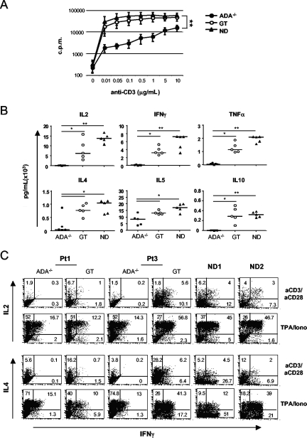
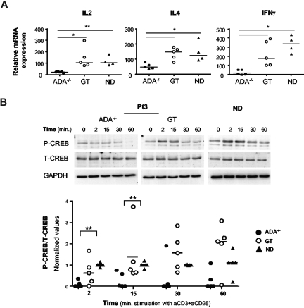
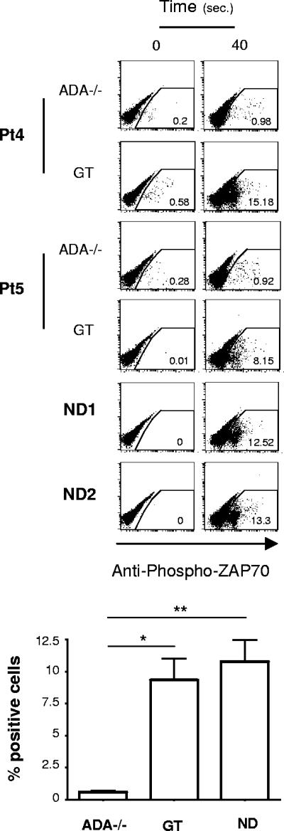
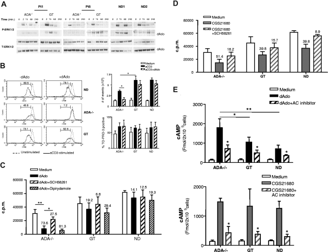
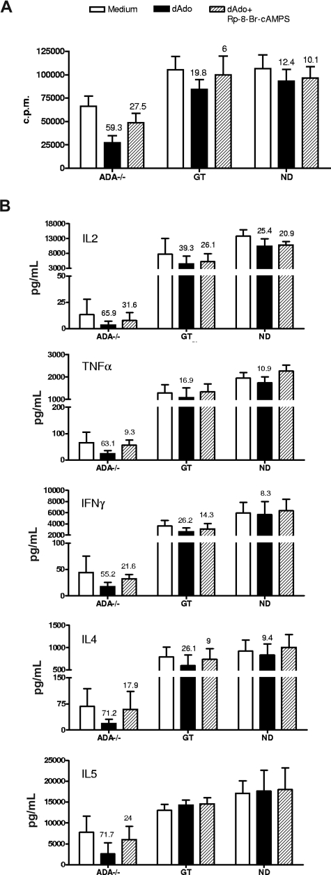
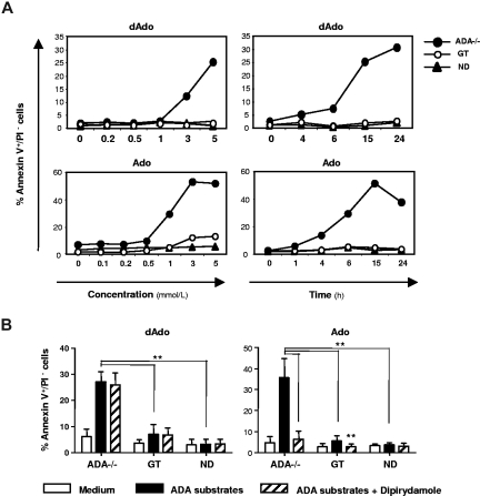
Mutations in the adenosine deaminase (ADA) gene are responsible for a form of severe combined immunodeficiency (SCID) caused by the lymphotoxic accumulation of ADA substrates, adenosine and 2'-deoxy-adenosine. The molecular mechanisms underlying T-cell dysfunction in humans remain to be elucidated. Here, we show that CD4(+) T cells from ADA-SCID patients have severely compromised TCR/CD28-driven proliferation and cytokine production, both at the transcriptional and protein levels. Such an impairment is associated with an intrinsically reduced ZAP-70 phosphorylation, Ca(2+) flux, and ERK1/2 signaling and to defective transcriptional events linked to CREB and NF-kappaB. Moreover, exposure to 2'-deoxy-adenosine results in a stronger inhibition of T-cell activation, mediated by the aberrant A(2A) adenosine receptor signaling engagement and PKA hyperactivation, or in a direct apoptotic effect at higher doses. Conversely, in T cells isolated from patients after gene therapy with retrovirally transduced hematopoietic stem/progenitor cells, the biochemical events after TCR triggering occur properly, leading to restored effector functions and normal sensitivity to apoptosis. Overall, our findings provide a better understanding of the pathogenesis of the immune defects associated with an altered purine metabolism and confirm that ADA gene transfer is an efficacious treatment for ADA-SCID. The trials in this study are enrolled at www.ClinicalTrials.gov as #NCT00598481 and #NCT0059978.

Trial registration: ClinicalTrials.gov NCT00598481 NCT00599781.


Figures

Figure 1
Figure 1
Characterization of the functional defect in CD4+ADA-deficient T cells. (A) Purified CD4+ T-cell lines from 5 patients and 5 NDs were stimulated with immobilized anti-CD3 mAb at the indicated concentrations, and [3H]thymidine incorporation was measured after 48 hours. One representative experiment of 3 is shown. (B) Cells were stimulated with 1 μg/mL anti-CD3, and culture supernatants were collected after 18 (IL-2) or 48 hours (all other cytokines). Cytokine concentrations were quantified by capture ELISA. Each value represents the mean of cytokine concentration measured in triplicate cultures for patients (n = 5; ADA−/− and GT) and healthy controls (n = 5). Median values are also reported. Background levels were subtracted. One determination of 3 is shown. (C) Production of IFN-γ vs either IL-2, or IL-4, was analyzed by intracytoplasmic staining in CD4+ T-cell lines as described in “Cytokine detection.” The experiment shown is representative of 5 independent experiments. Two ADA-SCID patients, before and after GT, and 2 NDs are reported. Range values corresponding to 5 patients and 7 NDs are indicated in Table 1.
Figure 2
Figure 2
A defective transcription of cytokine genes in ADA-deficient T cells associates with reduced phosphorylation of CREB. (A) Cytokine gene expression after TCR/CD28-mediated stimulation of CD4+ T-cell lines from 5 patients (ADA−/− and GT) and 4 NDs. Total RNA was prepared from either resting or stimulated cells (1 μg/mL anti-CD3 plus 10 μg/mL anti-CD28 mAbs) and gene expression were measured by semiquantitative RT-PCR. Values were normalized for the expression of HPRT and correspond to duplicate samples. Results are expressed as n-fold difference relative to resting condition. One representative experiment of 3 is shown. **P < .01; *P < .05. (B) Immunoblots depicting phosphorylated CREB, total CREB, and GAPDH. CD4+ T cells were stimulated with cross-linked (1 μg/mL), anti-CD3 plus (10 μg/mL), anti-CD28 mAbs for the indicated time points. The same membranes were sequentially probed for the different Abs. Upper panel: a representative experiment in Pt3 (ADA−/− and GT) and one ND is shown. Lower panel: densitometric analysis and statistical significance (**P < .01) on 5 patients (ADA−/− and GT) and 5 NDs are shown. Values corresponding to unstimulated cells were subtracted.
Figure 3
Figure 3
Defective proximal TCR signaling in ADA-deficient T cells. Intracytoplasmic analysis of phosphorylated ZAP-70 (Y319) in response to cross-linked (1 μg/mL), anti-CD3 plus (10 μg/mL), anti–CD28-mediated activation of CD4+ T cells. Dot plots from 2 ADA-SCID patients, before and after GT, and 2 NDs are reported. Results corresponding to frequency of positive cells after 40 seconds of stimulation, in one of 3 independent determinations, from 5 patients and 4 NDs are indicated in the graph. Values corresponding to basal phosphorylation were subtracted (*P < .05; **P < .01).
Figure 4
Figure 4
dAdo strongly inhibits ERKs activation and effector functions in ADA-deficient T cells activating A2A AR signaling. (A) ERK activation was analyzed in CD4+ cells stimulated with cross-linked (1 μg/mL), anti-CD3 plus (10 μg/mL), anti-CD28 mAbs in the absence or presence of dAdo (500 μM). Samples of untreated and dAdo-treated stimulated cells were prepared in parallel, run on different gels but probed with the different Abs at the same time. (B) CFSE-labeled CD4+ cells were cultured in plain medium or stimulated with immobilized anti-CD3 mAb (1 μg/mL) in the absence or presence of dAdo (500 μM). After 64 hours, cells were analyzed by flow cytometry. TO-PRO-3 was added at the time of FACS analysis. Histograms depicting the CFSE content of TO-PRO-3 viable cells are shown from one representative patient and one ND. The percentage of CFSEdim cells is indicated in each plot. Background levels corresponding to unstimulated cells were subtracted. The total number of trypan blue–negative viable cells and the frequency of TO-PRO-3–positive cells from CFSE/TO-PRO-3 dot plots of total events are shown for 5 patients and 4 NDs. (C) CD4+ cells stimulated with 1 μg/mL coated anti-CD3 mAb in the absence or presence of dAdo (500 μM) or (D) CGS21680 (100 μM). The specified compounds (SCH58261 or dipyridamole) were included 1 hour before the addition of dAdo. After 48 hours, cell proliferation was assessed by [3H]thymidine incorporation. The result of a representative experiment including 5 patients (ADA−/− and GT) and 5 NDs is reported. Percentages of inhibition with respect to the untreated condition are indicated. (E) Intracellular cAMP levels were determined in CD4+ T cells incubated for 15 minutes with culture medium containing 5 mM dAdo or 1 mM CGS21680. The inhibitor of adenylate cyclase (AC, 1 mM) was added 30 minutes before the other drugs. The data represent the mean plus or minus SD from 5 patients (ADA−/− and GT) and 4 NDs in one of 3 independent experiments. Statistics: **P < .01; *P < .05.
Figure 5
Figure 5
PKA hyperactivation is responsible for dAdo-mediated suppression of T-cell function in ADA-deficient cells. CD4+ T cells were stimulated with 1 μg/mL coated anti-CD3 mAb in the presence or absence of dAdo (500 μM) with or without the inhibitor of cAMP-dependent protein kinase A, Rp-8-Br-cAMPS (150 μM). (A) Rate of proliferation was evaluated by [3H]thymidine incorporation after 48 hours. (B) Culture supernatants were collected for cytokine production quantification by ELISA after 18 (IL-2) or 48 (all other cytokine) hours. Percentages of inhibition with respect to the untreated condition are indicated. The data represent the mean plus or minus SD of 5 patients (ADA−/− and GT) and 5 NDs from 1 of 3 independent experiments.
Figure 6
Figure 6
Dose- and time-dependent apoptosis induced in ADA-deficient T cells by ADA substrates. (A) Bulk T-cell lines were incubated with the indicated concentrations of purine metabolites and cultured for 15 hours (dAdo) or 6 hours (Ado). In time course experiments, T cells were incubated with dAdo (5 mM) or Ado (1 mM) for the indicated period. The percentages of early apoptotic cells (annexin V+/PI) are shown for Pt3 (ADA−/− and GT) and one ND as representative experiment. (B) Bulk T-cell lines from 5 patients (ADA−/− and GT) and 6 NDs were cultured for 15 hours with dAdo (5 mM) or 6 hours with Ado (1 mM) in the absence or presence of the inhibitor of nucleoside transporter, dipyridamole (1 μM). The percentages of annexin V+/PI cells as mean plus or minus SD are shown. Data are representative of at least 5 independent experiments (**P < .01).

Similar articles

Cited by

  • Immunogenetic Metabolomics Reveals Key Enzymes That Modulate CAR T-cell Metabolism and Function.
    Renauer P, Park JJ, Bai M, Acosta A, Lee WH, Lin GH, Zhang Y, Dai X, Wang G, Errami Y, Wu T, Clark P, Ye L, Yang Q, Chen S. Renauer P, et al. Cancer Immunol Res. 2023 Aug 3;11(8):1068-1084. doi: 10.1158/2326-6066.CIR-22-0565. Cancer Immunol Res. 2023. PMID: 37253111 Free PMC article.
  • A map of human circular RNAs in clinically relevant tissues.
    Maass PG, Glažar P, Memczak S, Dittmar G, Hollfinger I, Schreyer L, Sauer AV, Toka O, Aiuti A, Luft FC, Rajewsky N. Maass PG, et al. J Mol Med (Berl). 2017 Nov;95(11):1179-1189. doi: 10.1007/s00109-017-1582-9. Epub 2017 Aug 25. J Mol Med (Berl). 2017. PMID: 28842720 Free PMC article.
  • Defective B cell tolerance in adenosine deaminase deficiency is corrected by gene therapy.
    Sauer AV, Morbach H, Brigida I, Ng YS, Aiuti A, Meffre E. Sauer AV, et al. J Clin Invest. 2012 Jun;122(6):2141-52. doi: 10.1172/JCI61788. Epub 2012 May 24. J Clin Invest. 2012. PMID: 22622038 Free PMC article. Clinical Trial.
  • Early-onset stroke and vasculopathy associated with mutations in ADA2.
    Zhou Q, Yang D, Ombrello AK, Zavialov AV, Toro C, Zavialov AV, Stone DL, Chae JJ, Rosenzweig SD, Bishop K, Barron KS, Kuehn HS, Hoffmann P, Negro A, Tsai WL, Cowen EW, Pei W, Milner JD, Silvin C, Heller T, Chin DT, Patronas NJ, Barber JS, Lee CC, Wood GM, Ling A, Kelly SJ, Kleiner DE, Mullikin JC, Ganson NJ, Kong HH, Hambleton S, Candotti F, Quezado MM, Calvo KR, Alao H, Barham BK, Jones A, Meschia JF, Worrall BB, Kasner SE, Rich SS, Goldbach-Mansky R, Abinun M, Chalom E, Gotte AC, Punaro M, Pascual V, Verbsky JW, Torgerson TR, Singer NG, Gershon TR, Ozen S, Karadag O, Fleisher TA, Remmers EF, Burgess SM, Moir SL, Gadina M, Sood R, Hershfield MS, Boehm M, Kastner DL, Aksentijevich I. Zhou Q, et al. N Engl J Med. 2014 Mar 6;370(10):911-20. doi: 10.1056/NEJMoa1307361. Epub 2014 Feb 19. N Engl J Med. 2014. PMID: 24552284 Free PMC article.
  • Inhibition of ENT1 relieves intracellular adenosine-mediated T cell suppression in cancer.
    Sanders TJ, Nabel CS, Brouwer M, Hermant AL, Chaible L, Deglasse JP, Rosewick N, Pabois A, Cathou W, Smets A, Deligny M, Marchante J, Dubray Q, Letellier MC, Martinoli C, Marillier R, De Henau O, McGrath Y, Vander Heiden MG, Houthuys E. Sanders TJ, et al. Nat Immunol. 2025 Jun;26(6):854-865. doi: 10.1038/s41590-025-02153-3. Epub 2025 May 12. Nat Immunol. 2025. PMID: 40355731 Free PMC article.

References

    1. Giblett ER, Anderson JE, Cohen F, Pollara B, Meuwissen HJ. Adenosine-deaminase deficiency in two patients with severely impaired cellular immunity. Lancet. 1972;2:1067–1069. - PubMed
    1. Hirschhorn R, Roegner V, Rubinstein A, Papageorgiou P. Plasma deoxyadenosine, adenosine, and erythrocyte deoxyATP are elevated at birth in an adenosine deaminase-deficient child. J Clin Invest. 1980;65:768–771. - PMC - PubMed
    1. Buckley RH, Schiff RI, Schiff SE, et al. Human severe combined immunodeficiency: genetic, phenotypic, and functional diversity in one hundred eight infants. J Pediatr. 1997;130:378–387. - PubMed
    1. Hirschhorn R. Immunodeficiency disease due to deficiency of adenosine deaminase. In: Ochs H, Smith C, Puck J, editors. Primary Immunodeficiency Diseases. New York, NY: Oxford Univ. Press; 1999.
    1. Arredondo-Vega FX, Santisteban I, Daniels S, Toutain S, Hershfield MS. Adenosine deaminase deficiency: genotype-phenotype correlations based on expressed activity of 29 mutant alleles. Am J Hum Genet. 1998;63:1049–1059. - PMC - PubMed

Publication types

MeSH terms

Substances

Associated data