. 2008 Jun;22(6):1769-77.
doi: 10.1096/fj.07-087627. Epub 2008 Jan 24.

Fibroblast growth factor represses Smad-mediated myofibroblast activation in aortic valvular interstitial cells

Affiliations

Fibroblast growth factor represses Smad-mediated myofibroblast activation in aortic valvular interstitial cells

Melinda C Cushing et al. FASEB J. 2008 Jun.

Abstract

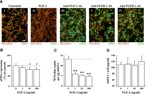
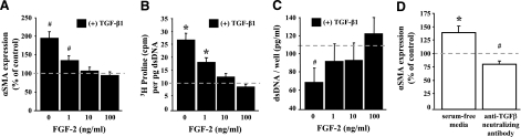
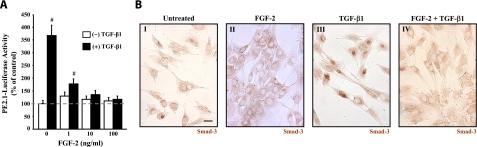
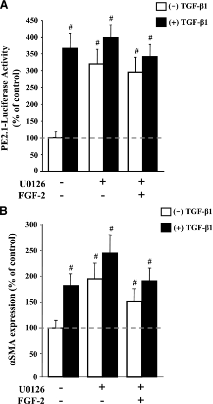
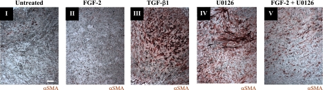
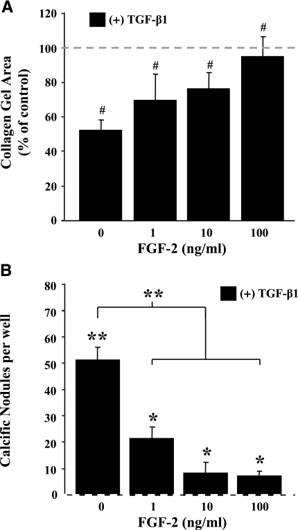
This study aimed to identify signaling pathways that oppose connective tissue fibrosis in the aortic valve. Using valvular interstitial cells (VICs) isolated from porcine aortic valve leaflets, we show that basic fibroblast growth factor (FGF-2) effectively blocks transforming growth factor-beta1 (TGF-beta1)-mediated myofibroblast activation. FGF-2 prevents the induction of alpha-smooth muscle actin (alphaSMA) expression and the exit of VICs from the cell cycle, both of which are hallmarks of myofibroblast activation. By blocking the activity of the Smad transcription factors that serve as the downstream nuclear effectors of TGF-beta1, FGF-2 treatment inhibits fibrosis in VICs. Using an exogenous Smad-responsive transcriptional promoter reporter, we show that Smad activity is repressed by FGF-2, likely an effect of the fact that FGF-2 treatment prevents the nuclear localization of Smads in these cells. This appears to be a direct effect of FGF signaling through mitogen-activated protein kinase (MAPK) cascades as the treatment of VICs with the MAPK/extracellular regulated kinase (MEK) inhibitor U0126 acted to induce fibrosis and blocked the ability of FGF-2 to inhibit TGF-beta1 signaling. Furthermore, FGF-2 treatment of VICs blocks the development of pathological contractile and calcifying phenotypes, suggesting that these pathways may be utilized in the engineering of effective treatments for valvular disease.


Figures

Figure 1
Figure 1
FGF-2 reduced markers of myofibroblast activation in VICs. A) Treatment of VICs with 10 ng/ml FGF-2 (II) reduced αSMA expression when compared with untreated control cell populations (I). Inhibition of FGF signaling with 50 μg/ml of antibodies that neutralize FGF-2 (III) or the FGF-2 receptors, FGFR-2 (IV) and FGFR-3 (V), caused a dramatic induction of de novo αSMA expression (green). Counterstaining of nucleic acid with propidium iodide is shown in red. All images were taken at the same magnification; scale bar = 10 μm. Previously published studies have demonstrated the efficacy of the anti-αSMA antibody to selectively stain stress fibers in myofibroblasts. B) Dose-dependent reduction of αSMA expression by FGF-2 in VICs. VICs were incubated for 48 h with FGF-2 at the concentrations indicated, and expression was detected by cell-based ELISA. Expression is shown as a percentage of the untreated (0 ng/ml) controls. C) Dose-dependent reduction in matrix deposition by FGF-2 in VICs. VICs were incubated for 72 h with FGF-2, and [3H]proline incorporation was measured by scintillation counter. [3H]proline levels were normalized to DNA content of each sample to account for potential differences in cell numbers between samples. D) Cell growth is unaffected by FGF-2. Subconfluent VIC cultures were treated with FGF-2 for 72 h, and the relative number of cells in each sample was determined by measuring total dsDNA content. In all charts, the dashed line indicates the level of untreated controls and represents the level to which treated samples are compared. #P < 0.05; **P < 0.001.
Figure 2
Figure 2
FGF-2 blocks TGF-β1-mediated myofibroblast activation in VICs. A) FGF-2 blocks the TGF-β1-mediated induction of αSMA expression. VICs were incubated for 48 h with 5 ng/ml TGF-β1 in combination with increasing concentrations of FGF-2, and αSMA expression was detected by cell-based ELISA. Expression is shown as a percentage of the untreated controls. B) FGF-2 blocks the TGF-β1-mediated induction of matrix deposition. VICs were incubated for 72 h with 5 ng/ml TGF-β1 in combination with increasing concentrations of FGF-2. [3H]proline incorporation was then measured by scintillation counter. [3H]proline levels were normalized to DNA content of each sample to account for potential differences in cell numbers between samples. C) FGF-2 blocks the TGF-β1-mediated reduction in cell growth. Subconfluent VIC cultures were treated with 5 ng/ml TGF-β1 in combination with increasing concentrations of FGF-2. After 72 h, the relative number of cells in each sample was determined by measuring total dsDNA content. Normal culturing conditions for VICs represses αSMA expression even though TGF-β signaling appears active. VICs were incubated for 48 h in either serum-free media or treated with an anti-TGF-β neutralizing antibody, and αSMA expression was detected by cell-based ELISA. Expression is shown as a percent of the controls cultured in 15% FBS. In all charts, the dashed line indicates the level of untreated controls (0 ng/ml TGF-β1 and FGF-2) and represents the level to which treated samples are compared. #P < 0.05; *P < 0.01.
Figure 3
Figure 3
TGF-β1-stimulated Smad activity is modulated by FGF-2. A) FGF-2 blocks TGF-β1-mediated transcriptional induction of Smad-responsive promoter. VICs transfected with the Smad-responsive luciferase reporter plasmid PE2.1 were treated with increasing concentrations of FGF-2 and luciferase activity was measured after 48 h. TGF-β1 (5 ng/ml) caused a 3.5-fold increase in luciferase activity when compared with untreated controls. This increase is markedly attenuated when FGF-2 (1–100 ng/ml) is codelivered with TGF-β1. Dashed line indicates level of untreated controls (0 ng/ml TGF-β1 and FGF-2). B) FGF-2 blocks TGF-β1-mediated Smad-3 nuclear accumulation in VICs. VICs were treated with FGF-2 (10 ng/ml; II), TGF-β1 (5 ng/ml; III), or both (IV). Representative micrographs show cellular localization of Smad-3 by indirect immunoperoxidase staining. VICs treated with TGF-β1 alone showed increased nuclear staining, indicating that Smad-3 had been translocated to the nucleus in these cells. FGF-2 treated cells have cytoplasmic staining with obvious occlusion of Smad-3 from the nucleus. All images were taken at the same magnification. Scale bar = 2.5 μm. #P < 0.05.
Figure 4
Figure 4
MAPK inhibitor blocks myofibroblast activation in VICs. A) The inhibition of ERK-1/2 prevents FGF-2 from blocking TGF-β1-medaited Smad activation. VICs were transfected with the Smad-responsive luciferase reporter plasmid PE2.1 and treated with a combination of the MEK-1/2 inhibitor U0126 (15 μmol/L), FGF-2 (10 ng/ml), and/or TGF-β1 (5 ng/ml). Luciferase activity was measured after 48 h after treatment. The MEK-1/2 inhibitor significantly increases Smad activity when compared with untreated controls. Moreover, U0126 prevented FGF-2 from reducing the TGF-β1-mediated activation of the Smad-responsive promoter that was observed in a previous experiment (Fig. 3A). B) The inhibition of ERK-1/2 prevents FGF-2 from blocking the TGF-β1-medaited induction of αSMA expression. VICs were treated with a combination of the MEK-1/2 inhibitor U0126 (15 mol/L), FGF-2 (10ng/ml), and/or TGF-β1 (5 ng/ml) for 48 h before αSMA detection by ELISA. The MEK-1/2 inhibitor U0126 significantly increased αSMA expression when compared with untreated controls. In addition, U0126 prevented FGF-2 from reducing the TGF-β1-mediated induction of αSMA that was observed in a previous experiment (Fig. 2A). In each chart, the dashed line indicates the level of untreated controls (0 ng/ml TGF-β1 and FGF-2) and represents the level to which treated samples are compared. #P < 0.05.
Figure 5
Figure 5
FGF-2 reduces fibrosis in intact aortic leaflets. αSMA expression in cultured porcine aortic valve leaflets is indicated by red immunoperoxidase staining. Intact valve leaflets were treated for 2 wk with control media (I), FGF-2 (10 ng/ml; II), TGF-β1 (5 ng/ml; III), U0126 (15 μmol/L; (IV), or U0126 (15 μmol/L) + FGF-2 (10 ng/ml; V). All images were taken at the same magnification, and the scale bar is equal to 20 μm. Previously published studies have demonstrated the efficacy of the anti-αSMA antibody to selectively stain stress fibers in myofibroblasts.
Figure 6
Figure 6
FGF-2 inhibits pathological phenotypes observed in TGF-β1-treated VICs. A) FGF-2 prevents fibrotic contraction of VICs treated with TGF-β1. VICs were encapsulated into 3-dimensional collagen matrices in 48-well tissue culture plates and treated with TGF-β1 (5 ng/ml) and FGF-2 (0–100 ng/ml) for 24 h. Matrices were then detached from the plate surface, and gel surface area was determined using ImageJ software. Collagen gel areas are plotted as a percentage of untreated control (0 ng/ml TGF-β1 and FGF-2). B) Dose-dependent reduction in TGF-β1-mediated calcific nodule formation by FGF-2 in VICs. VICs were seeded into 96-well plates and treated with TGF-β1 (5 ng/ml) and FGF-2 (0–100 ng/ml) for 72 h. The cells were then stained with Alizarin red to identify calcium-based mineralization, and the number of calcific nodules was counted in each well. No calcific nodules were observed in the untreated controls (0 ng/ml TGF-β1 and FGF-2), which are represented as a dashed line on the x axis. However, VICs treated with TGF-β1 showed a dramatic increase in nodule formation (>50 nodules/well), which is significantly attenuated by FGF-2 at concentrations ≥1 ng/ml. In each chart, the dashed line indicates the level of untreated controls (0 ng/ml TGF-β1 and FGF-2) and represents the level to which treated samples are compared, unless indicated by brackets. #P < 0.05; *P < 0.01; **P < 0.001.

Similar articles

Cited by

References

    1. Walker G. A., Masters K. S., Shah D. N., Anseth K. S., Leinwand L. A. Valvular myofibroblast activation by transforming growth factor-beta. Circ Res. 2004;95:253–260. - PubMed
    1. Kaden J. J., Kilic R., Sarikoc A., Hagl S., Lang S., Hoffmann U., Brueckmann M., Borggrefe M. Tumor necrosis factor alpha promotes an osteoblast-like phenotype in human aortic valve myofibroblasts: a potential regulatory mechanism of valvular calcification. Int J Mol Med. 2005;16:869–872. - PubMed
    1. Jian B., Narula N., Li Q. Y., Mohler E. R., 3rd, Levy R. J. Progression of aortic valve stenosis: TGF-beta1 is present in calcified aortic valve cusps and promotes aortic valve interstitial cell calcification via apoptosis. Ann Thorac Surg. 2003;75:457–465. - PubMed
    1. Desmouliere A., Redard M., Darby I., Gabbiani G. Apoptosis mediates the decrease in cellularity during the transition between granulation tissue and scar. Am J Pathol. 1995;146:56–66. - PMC - PubMed
    1. Rabkin-Aikawa E., Farber M., Aikawa M., Schoen F. J. Dynamic and reversible changes of interstitial cell phenotype during remodeling of cardiac valves. J Heart Valve Dis. 2004;13:841–847. - PubMed

Publication types

LinkOut - more resources