. 2008 Mar;9(3):293-300.
doi: 10.1038/sj.embor.7401161. Epub 2008 Jan 25.

NLRX1 is a mitochondrial NOD-like receptor that amplifies NF-kappaB and JNK pathways by inducing reactive oxygen species production

Affiliations

NLRX1 is a mitochondrial NOD-like receptor that amplifies NF-kappaB and JNK pathways by inducing reactive oxygen species production

Ivan Tattoli et al. EMBO Rep. 2008 Mar.

Abstract

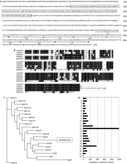
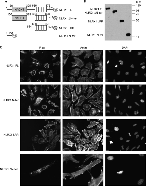
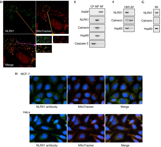
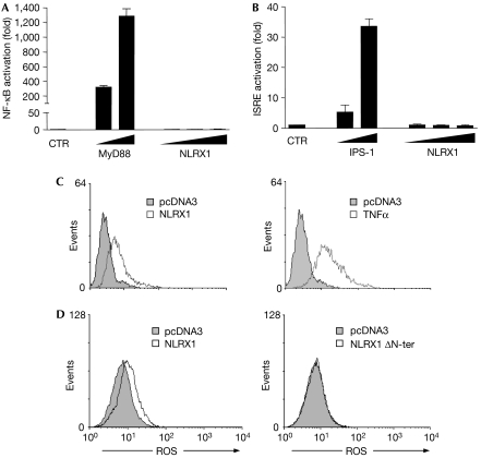
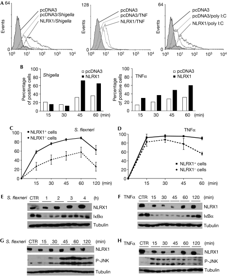
NOD-like receptors (NLRs) are a family of intracellular sensors of microbial- or danger-associated molecular patterns. Here, we report the identification of NLRX1, which is a new member of the NLR family that localizes to the mitochondria. NLRX1 alone failed to trigger most of the common signalling pathways, including nuclear factor-kappaB (NF)-kappaB- and type I interferon-dependent cascades, but could potently trigger the generation of reactive oxygen species (ROS). Importantly, NLRX1 synergistically potentiated ROS production induced by tumour necrosis factor alpha, Shigella infection and double-stranded RNA, resulting in amplified NF-kappaB-dependent and JUN amino-terminal kinases-dependent signalling. Together, these results identify NLRX1 as a NLR that contributes to the link between ROS generation at the mitochondria and innate immune responses.


Conflict of interest statement

The authors declare that they have no conflict of interest.

Figures

Figure 1
Figure 1
Characterization of NLRX1. (A) Amino-acid sequence of NLRX1. The NACHT domain is indicated by boxes, and the seven repeats of the LRR domain are numbered and indicated by arrows. (B) Sequence alignment of NLRX1 amino-terminal region from mouse (mNLRX1), rat (rNLRX1), Canis familiaris (cfNLRX1), human (hNLRX1) and Pan troglodytes (ptNLRX1). The residues identical and similar to those of NLRX1 are shown by reverse and dark highlighting, respectively. (C) The phylogenetic tree of NLR LRR domains was created using ClustalW and Neighbour-Joining/UPGMA method version 3.6a3 algorithms. (D) Expression profile of NLRX1 in a panel of 24 human tissues: (1) uterus; (2) thyroid; (3) thymus; (4) testis; (5) stomach; (6) spleen; (7) skin; (8) prostate; (9) pituitary; (10) pancreas; (11) ovary; (12) muscle; (13) mammary gland; (14), lymphocytes; (15) lymph nodes; (16) lung; (17) liver; (18) kidney; (19) intestine; (20) heart; (21) colon; (22) brain; (23) bone marrow; and (24) adrenal gland. NLR, NOD-like receptor.
Figure 2ac
Figure 2ac
NLRX1 is a mitochondria-associated protein. (A) Schematic representation of the NLRX1 constructs. (B) Expression profile of NLRX1 constructs determined by western blotting using a Flag antibody. (C) Immunofluorescence analysis of Flag-tagged NLRX1 constructs expressed in HeLa cells using a Flag antibody. Filamentous actin and nuclei were stained using rhodamin-conjugated phalloidin and DAPI, respectively.
Figure 2dh
Figure 2dh
(D) Confocal microscopy analysis of NLRX1-overexpressing HeLa cells, stained for NLRX1 (Flag antibody) and mitochondria (MitoTracker dye). (EG) Cellular fractions of NLRX1-Flag-transfected HEK293 cells were obtained by using specific lysis buffers and analysed by western blotting using antibodies against PARP (nuclear protein), Calnexin (endoplasmic reticulum protein), Hsp60 (mitochondrial protein) and caspase 3 (cytosolic protein). Stepwise fractionation shows that NLRX1 is present in (E) membrane, (F) heavy membrane and (G) mitochondrial fractions. (H) Mitochondrial localization of endogenous NLRX1 in MCF-7 and HeLa cells, using a rabbit polyclonal NLRX1 antibody and detection of mitochondria with MitoTracker. CF, cytosolic fraction; DAPI, 4,6-diamidino-2-phenylindole; HEK293, human embryonic kidney 293 cells; HM, heavy membrane fraction; HSP, heat shock protein; IM, isolated mitochondria; LM, light membrane fraction; MF; membrane fraction; NF, nuclear fraction; NLR, NOD-like receptor; PARP, poly (ADP-ribose) polymerase.
Figure 3
Figure 3
NLRX1 triggers the generation of ROS. (A,B) HEK293T epithelial cells were transfected with increasing amounts (10, 100 and 250 ng) of NLRX1 expression vector together with luciferase-reporter constructs of (A) NF-κB and (B) interferon-stimulated response element (ISRE). Overexpression of MyD88 (A) or IPS-1 (B) were used as positive controls. Data shown are the mean±s.e.m. of duplicates and are representative of three independent experiments. (C) HeLa cells were transfected overnight with pcDNA3 or NLRX1 vectors (left), or with pcDNA3 followed by stimulation for 1 h with 10 ng/ml TNFα (right). ROS production was measured by using a redox-sensitive dye (CM-H2DCFDA) on live cells, followed by flow cytometry analysis. (D) HeLa cells were transfected overnight with pcDNA3, NLRX1, NLRX1 ΔN-ter or NLRX1 N-ter vectors and ROS were measured as in (C). CTR, control; HEK293, human embryonic kidney 293 cells; NF-κB, nuclear factor-κB; NLR, NOD-like receptor; ROS, reactive oxygen species; TNFα, tumour necrosis factor α.
Figure 4
Figure 4
Effect of NLRX1 on signalling pathways triggered by TNFα and Shigella. (A) HeLa cells were transfected overnight with pcDNA3 or NLRX1 vectors and infected for 1 h with Shigella (left), or stimulated for 1 h with 10 ng/ml TNFα (middle) or 3 h with 10 μg/ml poly I:C (right). ROS production was measured by using a redox-sensitive dye (CM-H2DCFDA) on live cells, followed by flow cytometry analysis. (B) Time-course analysis of ROS production induced by Shigella (left) and TNFα (right) in conditions as in (A). (C,D) HeLa cells grown on glass coverslips were transfected overnight with NLRX1 expression vector and infected with (C) Shigella or (D) stimulated with 10 ng/ml TNFα for various times, as indicated. The nuclear translocation of NF-κB p65 subunit (as an indicator of the activation of the NF-κB pathway) was evaluated by immunofluorescence in cells overexpressing NLRX1 (NLRX1+) or not (NLRX1). Data shown are the mean±s.e.m. of three independent experiments, and for each condition and time point a minimum of 500 cells were counted. (EH) HEK293T cells were transfected overnight with NLRX1 expression vector or pcDNA3 and infected with (E,G) Shigella flexineri or (F,H) stimulated with 10 ng/ml TNFα for different time periods, as indicated, and extracts were immunoblotted with antibodies against (E,F) IκBα or (G,H) phospho-JNK. NLRX1 expression was assessed using an Flag antibody and tubulin was used as a control for protein levels. CM-H2DCFDA, 5-(and-6)-chloromethyl-2′,7′-dichlorodihydro-fluorescein diacetate, acetyl ester; CTR, control; HEK293, human embryonic kidney 293 cells; NF-κB, nuclear factor-κB; NLR, NOD-like receptor; poly I:C, polyinosinic:polycytidylic acid; ROS, reactive oxygen species; TNFα, tumour necrosis factor α.

References

    1. Akira S, Takeda K (2004) Toll-like receptor signalling. Nat Rev Immunol 4: 499–511 - PubMed
    1. Desagher S, Osen-Sand A, Nichols A, Eskes R, Montessuit S, Lauper S, Maundrell K, Antonsson B, Martinou JC (1999) Bid-induced conformational change of Bax is responsible for mitochondrial cytochrome c release during apoptosis. J Cell Biol 144: 891–901 - PMC - PubMed
    1. Fritz JH, Ferrero RL, Philpott DJ, Girardin SE (2006) Nod-like proteins in immunity, inflammation and disease. Nat Immunol 7: 1250–1257 - PubMed
    1. Girardin SE, Yaniv M (2001) A direct interaction between JNK1 and CrkII is critical for Rac1-induced JNK activation. EMBO J 20: 3437–3446 - PMC - PubMed
    1. Girardin SE et al. (2001) CARD4/Nod1 mediates NF-κB and JNK activation by invasive Shigella flexneri. EMBO Rep 2: 736–742 - PMC - PubMed

Publication types

MeSH terms