. 2008 Feb;118(2):526-33.
doi: 10.1172/JCI33813.

Erythropoietin deficiency decreases vascular stability in mice

Affiliations

Erythropoietin deficiency decreases vascular stability in mice

Jing Chen et al. J Clin Invest. 2008 Feb.

Abstract

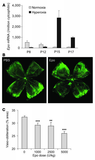
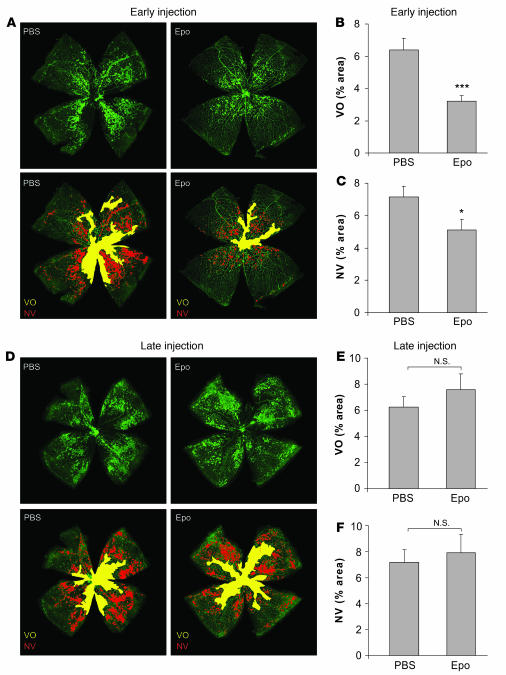
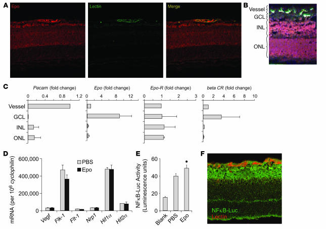
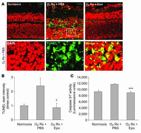
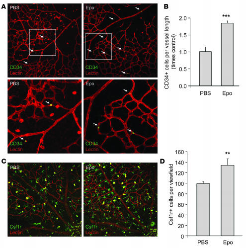
Erythropoietin (Epo), a hormone known to stimulate bone marrow erythrocyte production, is widely used to treat anemia in patients at risk for vascular disease. However, the effects of Epo on angiogenesis are not well defined. We studied the role of Epo in a mouse model of retinopathy characterized by oxygen-induced vascular loss followed by hypoxia-induced pathological neovascularization. Without treatment, local retinal Epo levels were suppressed during the vessel loss phase. Administration of exogenous Epo prevented both vessel dropout and subsequent hypoxia-induced neovascularization. Early use of Epo also protected against hypoxia-induced retinal neuron apoptosis. In contrast, retinal Epo mRNA levels were highly elevated during the retinopathy neovascular phase. Exogenous late Epo treatment did not protect the retina, but rather enhanced pathological neovascularization. Epo's early protective effect occurred through both systemic retinal recruitment of proangiogenic bone marrow-derived progenitor cells and activation of prosurvival NF-kappaB via Epo receptor activation on retinal vessels and neurons. Thus early retinal Epo suppression contributed to retinal vascular instability, and elevated Epo levels during the proliferation stage contributed to neovascularization and disease. Understanding the role of Epo in angiogenesis is critical to timing its intervention in patients with retinopathy or other diseases in which pathological angiogenesis plays a significant role.


Figures

Figure 1
Figure 1. Epo treatment prevents oxygen-induced retinal vessel loss in a dose-dependent manner.
(A) Real-time PCR quantification of Epo mRNA expression in age-matched mouse retinas under normoxia or hyperoxia treatment; copy number of Epo mRNA/106 copies of cyclophilin A control mRNA at P8, P12, P15, and P17 (n = 6 per group). (B) Representative retinal whole-mounts showing area of vaso-obliteration after 18 h of oxygen exposure and i.p. injection (P6 and P7) of Epo (right) or saline control (left). Original magnification, ×5. (C) Dose response of Epo protection against retinal vaso-obliteration at P8 (with i.p. injections at P6 and P7) (saline, n = 40; Epo 1,000 U/kg, n = 12; 2,500 U/kg, n = 7; 5,000 U/kg, n = 16) **P ≤ 0.01; ***P ≤ 0.001.
Figure 2
Figure 2. Early use of Epo protects against retinopathy with reduction in vaso-obliteration and neovascularization; in contrast, late use of Epo is not protective.
Retinas were whole-mounted at P17 after oxygen-induced retinopathy, showing both vaso-obliteration (VO) and neovascularization (NV). (A) Representative retina whole-mount with PBS control (left) or early Epo injections (right) (i.p., 5,000 U/kg, P6, P8, P10, and P12). Areas of vaso-obliteration (yellow) and neovascularization (red) were quantified. Original magnification, ×5. (B) Vaso-obliteration (***P ≤ 0.001) and (C) neovascularization (*P ≤ 0.02) in the mice with early Epo treatment at P17 (PBS, n = 40; Epo, n = 23). (D) Representative retina whole-mount with PBS control (left) or late Epo injections (right) (i.p., 5,000 U/kg, P14, P15, and P16) with areas of vaso-obliteration (yellow) and neovascularization (red). Original magnification, ×5. (E) Vaso-obliteration and (F) neovascularization in the mice with late Epo treatment (PBS, n = 34; Epo, n = 15).
Figure 4
Figure 4. Localization of Epo and Epo receptors and Epo-induced NF-κB activity in the retina.
(A) Representative retinal cross-sections from P8 normoxia mouse immunolabeled with Epo antibody (red) and lectin (green). (B) Dehydrated retinal cross-section from P8 normoxia retina stained with lectin (green) and counter-stained with H&E for laser capture microdissection. (C) mRNA expression of Pecam, Epo, Epo receptor (Epo-R), and β-common receptorCR) in laser-captured retinal cell layers (n = 6 per group). GCL, ganglion cell layer; INL, inner nuclear layer; ONL, outer nuclear layer. (D) Real-time PCR quantification of Vegf and Vegf receptor (Flk-1, Flt-1, Nrp1, Hif1a, and Hif2a) mRNA in retina of mouse littermate with Epo treatment (P6 and P7) or PBS control (n = 6 per group). Copy number of mRNA/106 copies cyclophilin A control mRNA were measured at P8 (n = 8 per group). (E) P8 oxygen-treated retinas from NF-κB–Luc reporter mice with PBS (n = 6) or Epo treatment (i.p., 5,000 U/kg, P6 and P7) (n = 7) showing NF-κB–Luc activity (*P ≤ 0.05). (F) Cross-section of P8 oxygen-treated retinas from NF-κB–Luc reporter mice stained with lectin GS-IB4 (red) and luciferase antibody (green) showing NF-κB localization. Original magnification, ×40.
Figure 3
Figure 3. Early Epo treatment protects retinal neurons from hypoxia-induced apoptosis.
(A) TUNEL (green) and DAPI (red) staining of retinal cross-sections from P17 normoxia mice and oxygen-treated mice with early Epo injection (i.p., 5,000 U/kg, at P6, P8, P10, and P12) or PBS control. A portion of the image from P17 oxygen-treated PBS control retina was enlarged (white frame) in lower panels. Original magnification, ×40. (B) Quantification of the fluorescence intensity of TUNEL stain in P17 normoxia mice and P17 hypoxia mice with Epo treatment or PBS control (n = 3 per group; *P ≤ 0.05). (C) Caspase-3/-7 activity in retinal homogenate isolated from P17 normoxia mice and P17 hypoxia mice with Epo treatment or PBS control (n = 6 per group; ***P ≤ 0.001).
Figure 5
Figure 5. Epo-induced increase of EPC and microglia numbers in the retina.
(A) Representative images of retinal flat mount from P8 oxygen-treated mice with Epo injection (i.p., 5,000 U/kg, P6 and P7) or PBS control stained with CD34 antibody (green) and lectin GS-IB4 (red), with a portion of the image enlarged (white frames). White arrows indicate CD34+ cells. (B) Mean number of EPCs (CD34+ cells) per vessel length in Epo treated compared with control retina (n = 6 per group; ***P ≤ 0.001). (C) Representative images of retinal flat mount from P8 oxygen-treated mice with Epo injection (i.p., 5,000 U/kg, P6 and P7) or PBS control stained with Csf-1R antibody (green) and lectin GS-IB4 (red). (D) Mean number of microglia (Csf-1R+ cells) in Epo-treated versus control retinas (n = 10 per group; **P ≤ 0.005). Original magnification, ×20.

Comment in

References

    1. Gariano R.F., Gardner T.W. Retinal angiogenesis in development and disease. Nature. 2005;438:960–966. - PubMed
    1. Sasaki R. Pleiotropic functions of erythropoietin. Intern. Med. 2003;42:142–149. - PubMed
    1. Katsura Y., et al. Erythropoietin is highly elevated in vitreous fluid of patients with proliferative diabetic retinopathy. Diabetes Care. 2005;28:2252–2254. - PubMed
    1. Watanabe D., et al. Erythropoietin as a retinal angiogenic factor in proliferative diabetic retinopathy. N. Engl. J. Med. 2005;353:782–792. - PubMed
    1. Singh A.K., et al. Correction of anemia with epoetin alfa in chronic kidney disease. N. Engl. J. Med. 2006;355:2085–2098. - PubMed

Publication types

MeSH terms