. 2008 Apr;129(4):463-78.
doi: 10.1007/s00418-008-0378-y. Epub 2008 Jan 25.

Early incorporation of obscurin into nascent sarcomeres: implication for myofibril assembly during cardiac myogenesis

Affiliations

Early incorporation of obscurin into nascent sarcomeres: implication for myofibril assembly during cardiac myogenesis

Andrei B Borisov et al. Histochem Cell Biol. 2008 Apr.

Abstract

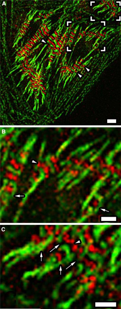
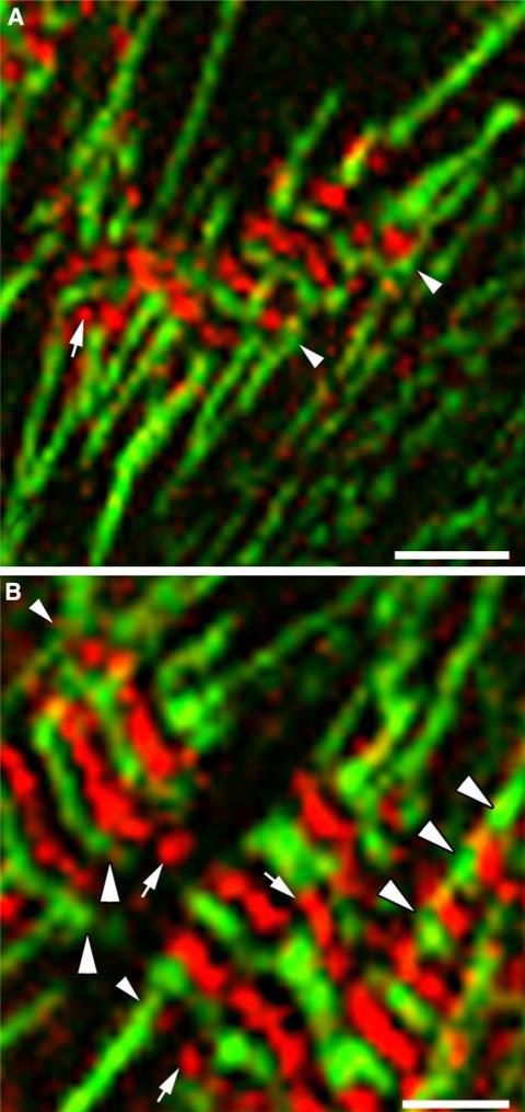
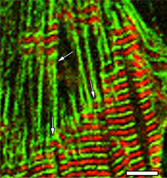
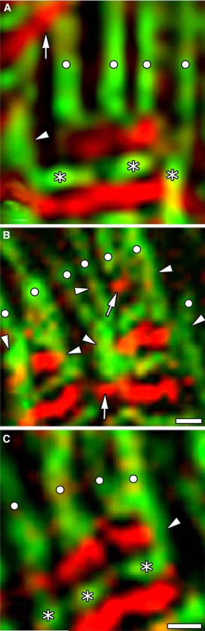
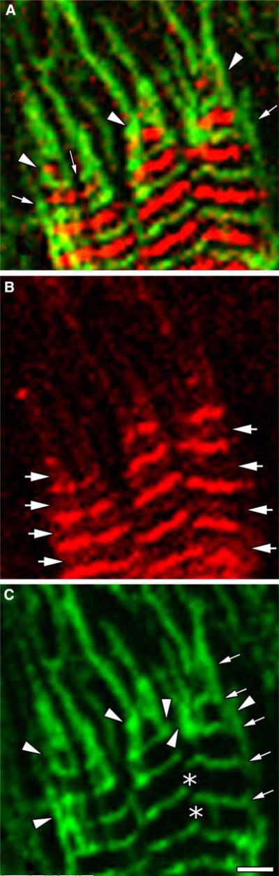
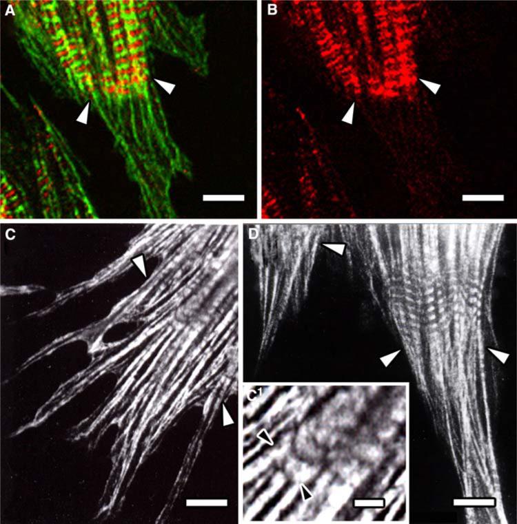
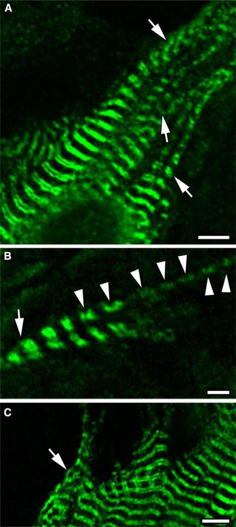
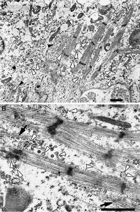
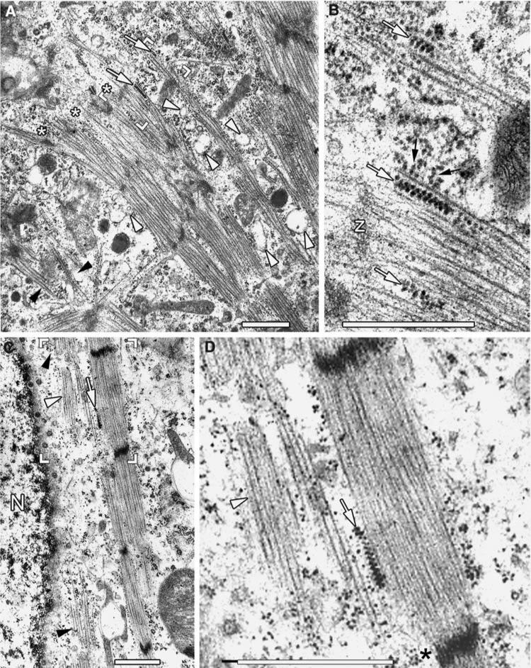
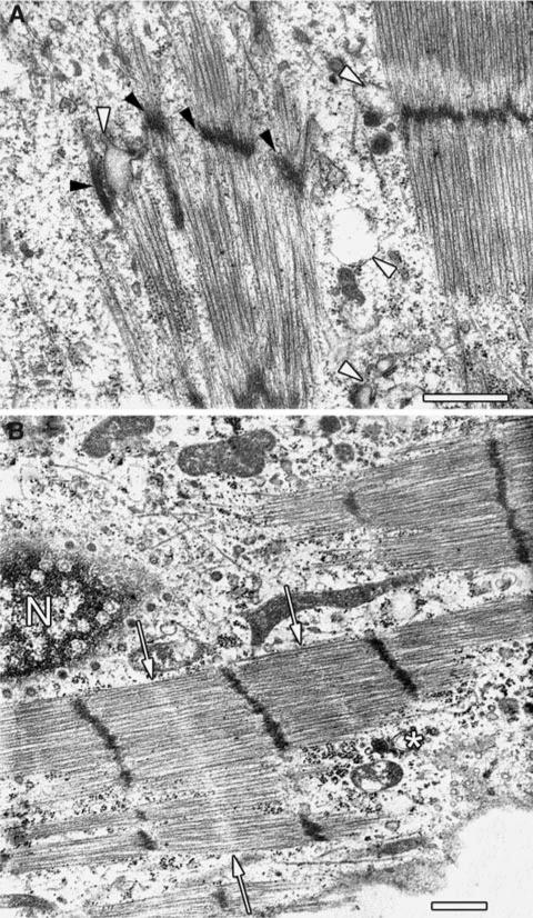
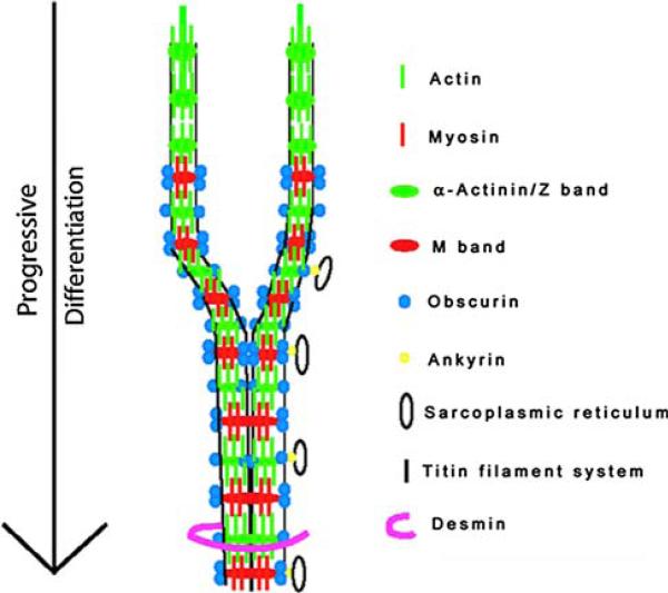
Obscurin is a recently identified giant multidomain muscle protein whose functions remain poorly understood. The goal of this study was to investigate the process of assembly of obscurin into nascent sarcomeres during the transition from non-striated myofibril precursors to striated structure of differentiating myofibrils in cell cultures of neonatal rat cardiac myocytes. Double immunofluorescent labeling and high resolution confocal microscopy demonstrated intense incorporation of obscurin in the areas of transition from non-striated to striated regions on the tips of developing myofibrils and at the sites of lateral fusion of nascent sarcomere bundles. We found that obscurin rapidly and precisely accumulated in the middle of the A-band regions of the terminal newly assembled half-sarcomeres in the zones of transition from the continuous, non-striated pattern of sarcomeric alpha-actinin distribution to cross-striated structure of laterally expanding nascent Z-discs. The striated pattern of obscurin typically ended at these points. This occurred before the assembly of morphologically differentiated terminal Z-discs of the assembling sarcomeres on the tips of growing myofibrils. The presence of obscurin in the areas of the terminal Z-discs of each new sarcomere was detected at the same time or shortly after complete assembly of sarcomeric structure. Many non-striated fibers with very low concentration of obscurin were already immunopositive for sarcomeric actin and myosin. This suggests that obscurin may serve for organization and alignment of myofilaments into the striated pattern. The comparison of obscurin and titin localization in these areas showed that obscurin assembly into the A-bands occurred soon after or concomitantly with incorporation of titin. Electron microscopy of growing myofibrils demonstrated intense formation and integration of myosin filaments into the "open" half-assembled sarcomeres in the areas of the terminal Z-I structures and at the lateral surfaces of newly formed, terminally located nascent sarcomeres. This process progressed before the assembly of the second-formed, terminal Z-discs of new sarcomeres and before the development of ultrastructurally detectable mature M-lines that define the completion of myofibril assembly, which supports the data of immunocytochemical study. Abundant non-aligned sarcomeres in immature myofibrils located on the growing tips were spatially separated and underwent the transition to the registered, aligned pattern. The sarcoplasmic reticulum, the organelle known to interact with obscurin, assembled around each new sarcomere. These results suggest that obscurin is directly involved in the proper positioning and alignment of myofilaments within nascent sarcomeres and in the establishment of the registered pattern of newly assembled myofibrils and the sarcoplasmic reticulum at advanced stages of myofibrillogenesis.


Figures

Fig. 1
Fig. 1
Obscurin incorporation during formation of individual myofibrillar clusters. a Assembling myofibrillar clusters in a cardiac muscle cell immunolabeled for sarcomeric α-actinin (green fluorescence) and obscurin (red fluorescence) shown at a low magnification. Immunolabeling for obscurin is observed as a striated pattern of myofibrils directly connected to non-striated continuous fibers intensely immuno-positive for the sarcomeric isoform of α-actinin. Note nearly complete absence of obscurin in the structures expressing the dotted and dashed patterns of α-actinin distribution. The frame corners indicate the areas shown in panel b (the lower set of corners) and c (the upper set of corners) at a high magnification. b The localization of sarcomeric α-actinin (green) and obscurin (red) in early myofibrils following formation of the first 2–3 differentiated sarcomeres. Note the presence of non-striated fibers (green) bifurcating from a nascent myofibril. The arrows show transiently preserved longitudinally oriented regions of α-actinin-positive non-striated fibers on each lateral side of the terminal sarcomeres on the tips of growing myofibrils at the areas of transition from the non-striated to striated pattern. Obscurin in such areas is located within the square or rectangular “boxes” immunopositive for α-actinin. The arrowhead shows obscurin-containing rounded bodies that assemble in the middle of nascent A-bands. c Lateral fusion of nascent myofibrils into a bigger cluster. Note the presence of non-striated fibers originating from the striated regions and bifurcating on the tips. The arrows show that longitudinally oriented elements of non-striated pattern on the lateral surfaces of the nascent sarcomeres are still well preserved at the time of obscurin accumulation in the middle of the A-bands. Long thin arrows show the gaps in the structure of laterally fusing elements of the second-formed Z-discs. Note the presence of obscurin in the mid-A-bands in the same sarcomeres. The arrowhead indicates the beaded pattern of obscurin localization. Note the presence of closely apposed obscurin bodies. Day 4 in culture. Bars 4.3 μm (A), 2 μm (b, c)
Fig. 2
Fig. 2
Progressive transverse and longitudinal expansion of fusing myofibrillar clusters. a The presence of longitudinally oriented α-actinin-containing material at the sides of the peripheral obscurin-positive sarcomeres in expanding myofibrillar clusters (arrowheads). The arrow shows the location of a rounded obscurin-positive body in the terminal sarcomere before the formation of the second transversely oriented Z-disc. Note that non-striated fibers are longitudinally oriented on the left and the right sides of the area marked with the arrow. b Presence of obscurin at the periphery of laterally aligning myofibrils (the arrow on the right) and close to the surface outside of the forming myofibril cluster (the arrows on the left). The areas of transition from myofibrils to non-striated fibrils are marked with the small arrowheads. Note the presence of obscurin in the A-bands, poor differentiation of the second-formed Z-discs of terminal sarcomeres in these areas, and different width and length of the Z-discs in newly formed myofibrils. Large arrowheads mark the periodicity of nascent Z-disc material in newly formed sarcomeres containing obscurin (the left side of the photo) and in more mature sarcomeres (the lower right side of the photo). Note increasing spacing in more mature sarcomeres compared to the terminal sarcomere. Bars 4 μm (a), 2 μm (b)
Fig. 3
Fig. 3
Obscurin incorporation (red fluorescence) into the contractile apparatus of a cardiac myocyte at the stage of formation of well-developed and laterally aligned myofibrillar clusters after 7 days in culture. myofibrils are labeled with the antibody to sarcomeric α-actinin (green fluorescence). The arrows show the sites of early incorporation of obscurin in the zones of lateral fusion of non-striated continuous fibers immunopositive for sarcomeric α-actinin. Note the presence of obscurin in the gaps between two fusing fibers and formation of the elements of nascent Z-discs in the zone of transition from the continuous pattern to sarcomeric cross-striations below and above obscurin-positive spots. Also note a progressive increase in sarcomere length in more mature regions of the same myofibrils (the lower part of the photo). Bar 5 μm
Fig. 4
Fig. 4
Patterns of obscurin (red fluorescence) and sarcomeric α-actinin (green fluorescence) localization in the areas of transition from non-striated fibrillar structures to cross-striated nascent myofibrils. a Non-striated fibrils continuously labeled for sarcomeric α-actinin are marked with white circles. The asterisks show the localization of the first-formed transversely oriented nascent Z-discs in each nascent myofibril. The arrowhead shows the preservation of a non-striated region of a longitudinally oriented continuously labeled fiber directly connected to a newly assembled Z-disc. Nascent Z-discs forming from individual non-striated fibers are still spatially separated from one another. The arrow indicates two closely opposed elongated, obliquely oriented obscurin-positive bodies located in the neighboring nascent myofibrils. Note that the edges of the obscurin-positive bodies extrude outside of the contour of a non-striated structure labeled with the white circle. The presence of both obscurin and α-actinin in the lateral surface of the non-striated fiber visible on the right side of the photo below the asterisk as yellow fluorescence of superimposing green and red labels. A clear zone free of obscurin is seen below the termination points of non-striated fibers on the left side of the photo above the Z-disc. b The process of lateral fusion of two nascent myofibrillar bundles. The symbols are the same is in panel a. Obscurin (red) starts to concentrate in the prospective A-band region before or at the beginning of lateral fusion of non-striated bundles (green) just after initial formation of the first Z-discs of each new sarcomere (the arrowheads and the arrow, on the top of the photo). Note that after disassembly of the components of non-striated fibers on the lateral surfaces of nascent sarcomeres, the fusion of laterally growing obscurin bands forms an uninterrupted striated pattern in the middle of A-bands (the arrow at the bottom). c advanced stages of Z-disc formation. The symbols are the same as in a. Note that the second Z-discs in nascent myofibrils are assembled following incorporation of obscurin and formation of clear spaces for the A-band assembly. Note the morphological difference of the obscurin-positive bodies (red) spatially separated in nascent sarcomeres and laterally fused A-bands in more mature sarcomeres located below. Bars 0.9 μm
Fig. 5
Fig. 5
Localization of obscurin (red fluorescence) and α-actinin (green fluorescence) in the terminal sarcomeres of a growing myofibrillar cluster. a merged image showing the localization of both proteins. The arrowheads show the preservation of elements of non-striated fibrils on the lateral surfaces of newly formed early sarcomeres at the sites of obscurin incorporation. Thin long arrow shows the gap in the nascent Z-band at the site of lateral fusion of two early myofibrillar bundles. Short arrows show transient presence of longitudinally oriented non-striated structures contacting Z-discs of newly formed sarcomeres. b The localization of obscurin in the same field. The arrows show the presence of obscurin in the Z-discs. Note that obscurin incorporates first in the area of nascent A-bands of each terminal sarcomere. Also note the absence of obscurin on the lateral surfaces of nascent sarcomeres in the areas immunopositive for sarcomeric α-actinin (see c). c Localization of α-actinin in the same field. The arrowheads indicate the presence of α-actinin on the lateral surfaces of the nascent sarcomeres on the tips of growing myofibrils. The arrows on the right show Z-disc periodicity along growing myofibrils. The asterisks show the gaps in nascent Z-discs that are located to newly formed myofibrils. The gap is nearly closed in the Z-bands located below the lateral fusion point. As one can see in photo b, obscurin bands in this area extrude to the periphery of sarcomeres and closely approach one another. Note a short distance between two nascent Z-discs in the terminal sarcomere at the initial stage of obscurin incorporation. Bar 1.8 μm
Fig. 6
Fig. 6
a Double immunofluorescent labeling of sarcomeric α-actinin (green fluorescence) and obscurin (red fluorescence) at the site of myofibrillogenesis in the peripheral cytoplasmic process of a cardiac muscle cell. b The localization of obscurin in the same field. Labeling of sarcomeric myosin heavy chains and sarcomeric actin in the areas of nascent myofibril formation at similar locations is shown in panels c, c1 and d, respectively. The arrowheads in ad show the zone of transition of growing myofibril clusters into non-striated structural precursors of myofibrils. Note the presence of the sarcomeric isoforms of α-actinin, myosin, and actin in continuously labeled non-striated fibers and the abrupt termination of intense obscurin labeling in the zones of transition from the striated to non-striated pattern. c1 Shows the presence of sarcomeric myosin both in the newly formed sarcomeres and in continuously labeled non-striated structures extending from assembling myofibrils. Bars 6 μm (a, b), 2 μm (c1), 10 μm (c, d)
Fig. 7
Fig. 7
Localization of titin in the growing myofibrillar clusters. a A general view of a cardiac muscle cell after 7 days in culture in the area of transition from the differentiated and aligned myofibrils to nascent myofibrils arrows show the points of bifurcation and trifurcation of nascent myofibrillar bundles. b A bifurcating growing myofibril shown at a higher magnification. The arrow shows the point of bifurcation. The arrowheads indicate the presence of periodically arranged immunopositivity for titin in nascent sarcomeres beyond the point of bifurcation. c Uninterrupted cross-striated titin pattern illustrating complete lateral alignment of myofibrils in a muscle cell after 12 days in culture. Note that the gaps between individual myofibril bundles are closed. The arrow shows a cytoplasmic ruZe with continuing myofibrollogenesis and spot-like periodic pattern of titin localization. Bars 4.2 μm (a), 1.7 μm (b), and 3.8 μm (c)
Fig. 8
Fig. 8
Early stage of formation of a myofibril cluster. a Nascent myofibrils are spatially separated from one another, but their Z-discs are aligned at approximately the same level. White arrowheads show the developing sarcoplasmic reticulum located closely to the lateral surfaces of myofibrils. White arrows show the presence of protein material connecting Z-lines at the neighboring myofibrils. Black arrowheads show free myosin filaments located close to the sites of assembly of nascent sarcomeres. b Branching myofibrils. The arrowheads indicate transversely and obliquely sectioned membranous tubular structures. Some of these structures represent the developing sarcoplasmic reticulum and are located closely to newly formed sarcomeres. White arrows show electron-dense protein deposits located near the lateral surfaces of Z-discs. Black arrows indicate the location of free myosin filaments. The asterisk shows myosin filaments located at angles to one another in a nascent terminal sarcomere. Four days in culture. Bars 1 μm
Fig. 9
Fig. 9
Electron microscopic images of myofibril assembly in rat cardiac myocytes. a Clusters of nascent myofibrils. Immature precursors of Z-discs are spatially separated and poorly aligned in relation to one another. Note that myosin filaments are loosely arranged and are oriented at different angles in the incompletely assembled terminal sarcomeres on the tips of growing myofibrils (asterisks). Also note the presence of transversely and obliquely sectioned cisterns of the forming sarcoplasmic reticulum (white arrowheads) located closely to the sites of myofibril assembly. Black arrowheads indicate free myosin filaments oriented at different angles and located closely to a nascent Z-disc. The arrows show the localization of polyribosomes on the tips of growing myofilaments of terminal sarcomeres. The frame corners show the area enlarged in panel b. The arrows in b show the same locations as in a at a higher magnification. b White arrows show the localization of polyribosomes attached to elongating sarcomeric filaments. Black arrows indicate free small spiral polysomes located closely to myosin filaments on the lateral surface of the forming sarcomere. A nascent Z-disc at early stage of formation is marked with the letter z. Note the presence of free growing sarcomeric filaments in the upper part of the photograph. c Nascent myofibrils in the perinuclear area. N marks the nucleus. Electron lucent zones in the middle of newly formed A-bands show the absence of M-lines (also see d). Arrowheads show the groups of poorly aligned free myosin filaments. The arrow shows a polyribosome oriented along myosin filaments in the A-band and located on the lateral surface of the sarcomere. d A high magnification view of the sarcomere, free myosin filaments and the polyribosome marked with frame corners in photo c. The arrow and white arrowhead show the same locations as in photo c. Note the absence of Z-discs between the groups of longitudinally oriented myosin filaments of different length located on the left side of the photo and marked with the arrowhead. Also note that the Z-disc and the A-band located on the top of the photo are wider than the Z-disc and the A-band of the same sarcomere at the bottom located closer to growing tip of the myofibril. The presence of a polyribosome in the narrowing area of A-band at a close distance from the narrow region of the terminal Z-disc marked with the asterisk shows that the longitudinal and lateral growth of the second Z-disc occurs following the assembly of myosin filament and more or less complete formation of the A-bands in nascent myofibrils. Also note the absence of M-line in the middle of the A-band. Bars 1 μm
Fig. 10
Fig. 10
Non-aligned and aligned nascent myofibrils. a Non-aligned myosin filaments and spatially separated Z-discs (black arrowheads) in immature sarcomeres. Note that the nascent Z-discs are narrow, spatially separated, elongated, and obliquely oriented. The peripheral Z-discs are structurally associated with actin and nascent myosin filament forming the I–Z–I structures. A sarcomere at more advanced stage of differentiation is shown on the right. Note the presence of transversely oriented Z-disc and longitudinally aligned myosin filaments organized in a clearly visible A-band. White arrowheads show the elements of the developing sarcoplasmic reticulum. b Lateral alignment of smaller myofibrils into a larger myofibril. The arrows indicate the electron-lucent zones in the middle of A-bands, which shows the absence of M-lines. The asterisk shows the localization of the transversely sectioned membranous tubular structure morphologically resembling a forming element of the T-tubule system. Bars 1 μm
Fig. 11
Fig. 11
The hypothetic model illustrating the functional role of obscurin during myofibril assembly that illustrates the mechanism of maturation of myofibrils and the role of obscurin in this process Currently available data suggest that obscurin plays a role of a “stitching” linker protein involved in binding of sarcomeric filaments and the establishment of the longitudinal alignment of nascent myofibrils in relation to one another and to the sarcoplasmic reticulum

Similar articles

Cited by

References

    1. Agarkova I, Perriard J-C. The M-band: an elastic web that cross-links thick filaments in the center of the sarcomere. Trends Cell Biol. 2005;15:477–485. - PubMed
    1. Agarkova I, Ehler E, Lange S, Schoenauer R, Perriard JC. M-band: a safeguard for sarcomeric stability? J Muscle Res Cell Motil. 2003;24:191–203. - PubMed
    1. Anversa P, Olivetti G, Bracchi PG, Loud AV. Postnatal development of the M-band in rat cardiac myofibrils. Circ Res. 1981;48:561–568. - PubMed
    1. Armani A, Galli S, Giacomello E, Bagnato P, Barone V, Rossi D, Sorrentino V. Molecular interactions with obscurin are involved in the localization of muscle-specific small ankyrin 1 isoforms to subcompartments of the sarcoplasmic reticulum. Exp Cell Res. 2006;312:3456–3458. - PubMed
    1. Arimura T, Matsumoto Y, Okazaki O, Hayashi T, Takahashi M, Inagaki N, Hinohara K, Ashizawa N, Yano K, Kimura A. Structural analysis of obscurin gene in hypertrophic cardiomyopathy. Biochem Biophys Res Commun. 2007;362:281–287. - PubMed

Publication types

Substances

LinkOut - more resources