

Review
doi: 10.1002/jmri.21263.
Stem cell therapy: MRI guidance and monitoring
Affiliations
- PMID: 18219684
- PMCID: PMC3075622
- DOI: 10.1002/jmri.21263
Item in Clipboard
Review
Stem cell therapy: MRI guidance and monitoring
J Magn Reson Imaging.
2008 Feb.
Abstract
With the recent advances in magnetic resonance (MR) labeling of cellular therapeutics, it is natural that interventional MRI techniques for targeting would be developed. This review provides an overview of the current methods of stem cell labeling and the challenges that are created with respect to interventional MRI administration. In particular, stem cell therapies will require specialized, MR-compatible devices as well as integration of graphical user interfaces with pulse sequences designed for interactive, real-time delivery in many organs. Specific applications that are being developed will be reviewed as well as strategies for future translation to the clinical realm.
(Copyright) 2008 Wiley-Liss, Inc.
Figures
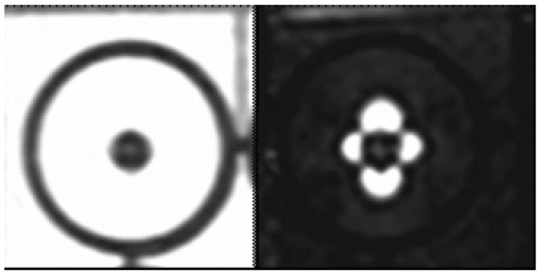
Fast spin echo image of one million iron oxide-labeled mesenchymal stem cells, which appear hypointense in an agarose phantom (left). Using a positive contrast imaging technique (25) the iron oxide-labeled stem cells appear hyperintense in a typical dipole pattern (right).
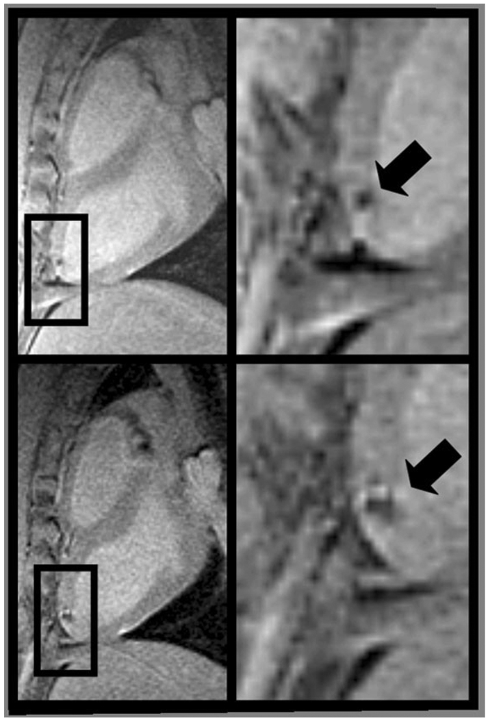
Long-axis MR images (left) of the left ventricle with magnified view (right) showing hypointense lesions (arrow) caused by iron oxide-labeled mesenchymal stem cells injected under x-ray fluoroscopy acquired within 24 hours (top) and 1 week (bottom) of injection. Expansion of the hypointense region at 1 week is indicative of local migration of the stem cells. Adapted from Kraitchman et al (38), which contains expanded contiguous image data and histological validation.
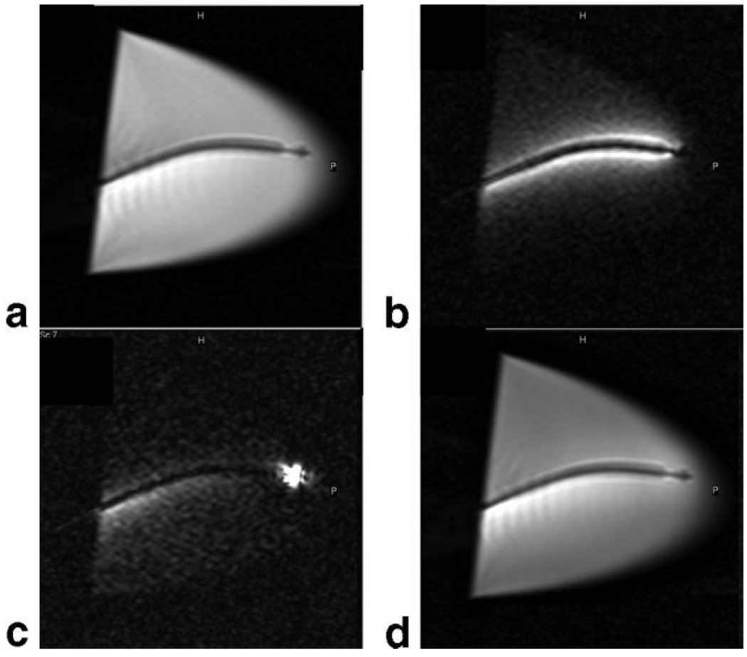
Long-axis MR images (4.0 msec repetition time, 2.0 msec echo time, 70° flip angle, 240 mm2 field of view, 160 × 160 matrix, 8 mm section thickness, 2 frames per second) acquired in water bath containing nitinol catheter and different active coil elements. a: Only the external surface coil elements were active. b: Only the catheter coil was active. c: Only the catheter tip microcoil was active. d: Both surface coil elements and active catheter coil are contributing to the image. Reprinted with permission from Saeed et al (65).
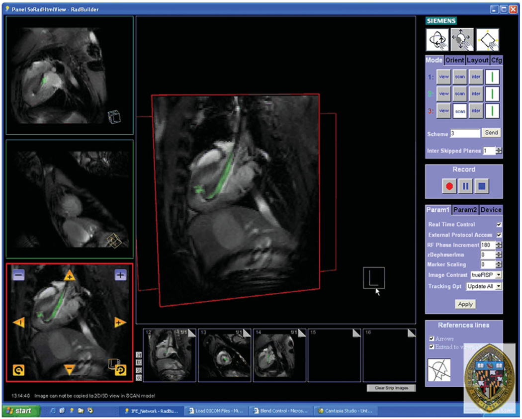
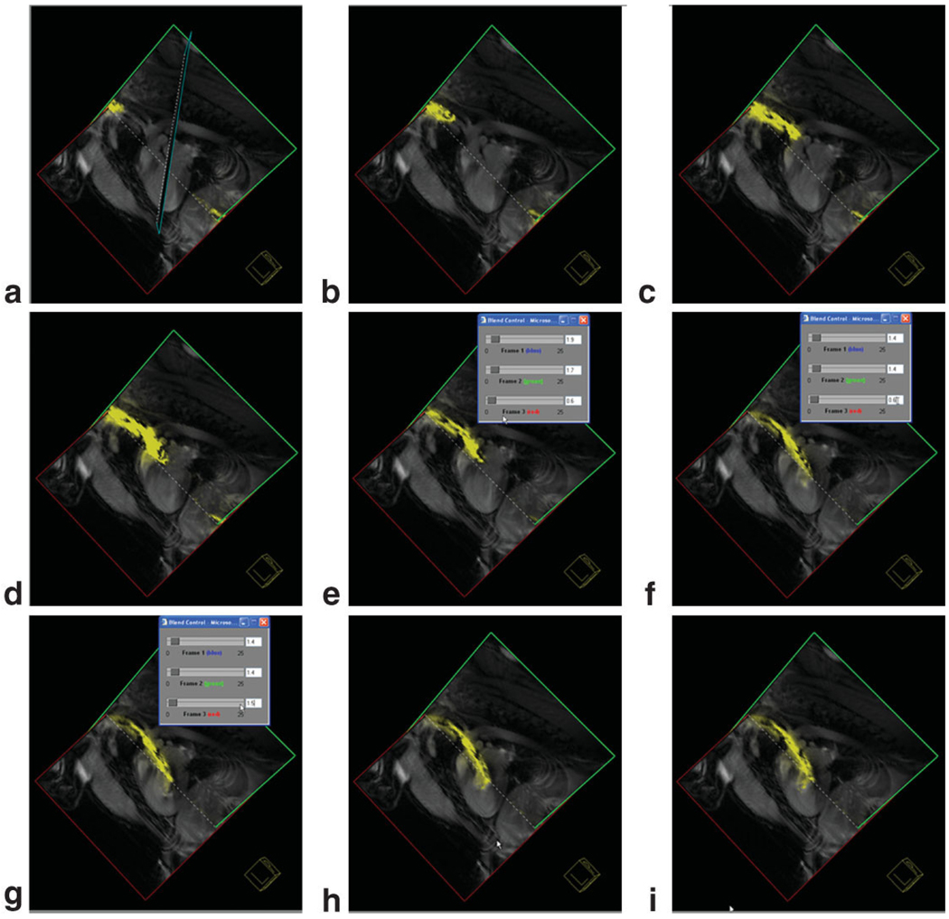
Screen capture of the Siemens prototype Interactive Front End (IFE) graphical interface that enables real-time scan plane manipulation and serial acquisition of up to three imaging planes. The image is reconstructed with the active injection catheter colored green for enhanced visibility. Representative pseudo long- and short-axis images are shown acquired in real-time in vivo in a canine reperfused myocardial infarction. Bookmark images (small images at bottom) facilitate rapid return to previous scan plane position using a simple drag-n-drop of the image plane into one of three image acquisition planes.
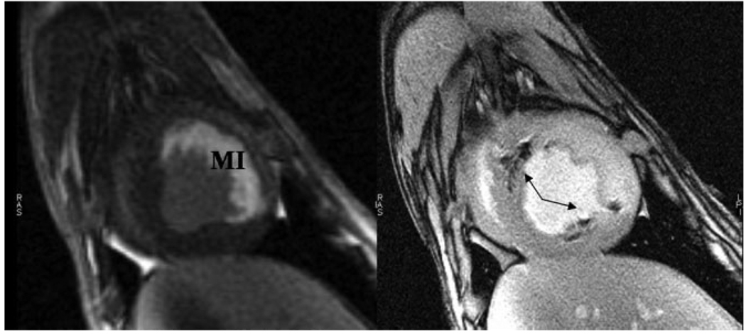
High-resolution, ECG-gated, breath-hold steady-state free precession short-axis images prior to contrast injection (left), and after transmyocardial gadolinium-based contrast injection with tissue vital dye (middle) under MR fluoroscopy. Postmortem digital image (right) demonstrates a high concordance of the spatial location and extent with in vivo MRI.
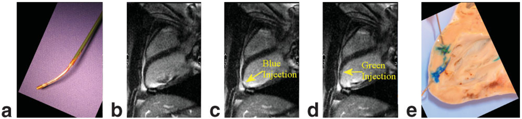
a: The distal tip of a custom, MR-compatible active injection catheter (66) that was used for injections of 10% gadolinium with a tissue vital dye. b: ECG-gated, breath-hold long-axis image prior to injection. c: Long-axis image after first gadolinium injection mixed with a blue dye. d: Long-axis image after second gadolinium injection with green dye. Unfortunately, injection sites can only be appreciated for a short period of time due to wash-out of the gadolinium contrast agent. e: Postmortem image demonstrating distinct injection sites. Adapted from Karmarkar et al (66).
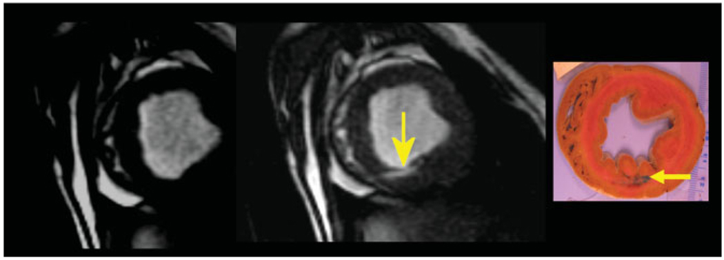
Targeting of the iron oxide-labeled stem cell injections to the peri-infarction area was performed based on delayed contrast-enhanced short-axis MRI (left) in which hyperintense signal represents myocardial infarction (MI) in this acute, reperfused canine model. Short-axis, high-resolution fast gradient echo image (right) of the left ventricle demonstrating multiple hypointensities (arrows) from iron oxide-labeled mesenchymal stem cells that were injected under MR fluoroscopy. Adapted from Bulte and Kraitchman (17).
Still-frame captures of three-plane view from the Siemens interactive graphical interface demonstrating guiding an active catheter into the left ventricle from a carotid artery approach. Images were acquired with a nongated steady-state free precession pulse sequence. The needle of the injection catheter is colored yellow. In frame (f) the gain from the active catheter is reduced to enable better determination of the catheter position.
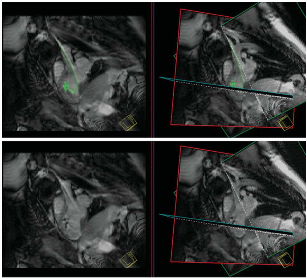
Top: A single pseudo-long axis-plane (left) and three-plane view (right) with an injection catheter shown in green prior to labeled stem cell injection. The catheter is steerable and flexible to enable access to many portions of the left ventricular endocardial surface. Bottom: During injection of iron oxide-labeled stem cells the active catheter gain is no longer colored to enhance detection of hypointensities in the myocardium to document stem cell injection success.
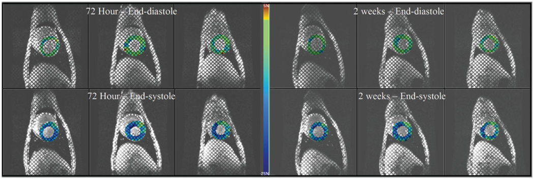
Three representative short-axis images at end-diastole (top row) and end-systole (bottom row) in a dog with a reperfused left anterior descending coronary artery myocardial infarction that received transmyocardially administered mesenchymal stem cells under MR fluoroscopy. Prior to injection at 72 hours postinfarction (images on left), tagged MRI with circumferential strain shown as a color overlay where less shortening is green and more shortening is blue demonstrates a mild functional defect in the anteroseptal wall (12 o’clock to 2 o’clock) that shows little functional improvement or slight worsening of function at 2 weeks posttransmyocardial mesenchymal stem cell delivery (images on right).
Similar articles
-
Pulse sequences and system interfaces for interventional and real-time MRI.J Magn Reson Imaging. 2008 Feb;27(2):267-75. doi: 10.1002/jmri.21268. J Magn Reson Imaging. 2008. PMID: 18219681 Review.
-
[Interventional MR imaging: state of the art and technological advances].J Radiol. 2008 Jan;89(1 Pt 1):13-20. doi: 10.1016/s0221-0363(08)70365-2. J Radiol. 2008. PMID: 18288022 Review. French.
-
MR-guided endovascular interventions: a comprehensive review on techniques and applications.Eur Radiol. 2008 Apr;18(4):645-57. doi: 10.1007/s00330-007-0818-4. Epub 2007 Dec 11. Eur Radiol. 2008. PMID: 18071710 Review.
-
Intracoronary injection of contrast media maps the territory of the coronary artery: an MRI technique for assessing the effects of locally delivered angiogenic therapies.Acad Radiol. 2008 Nov;15(11):1354-9. doi: 10.1016/j.acra.2008.09.002. Acad Radiol. 2008. PMID: 18995187
-
Targeted drug delivery under MRI guidance.J Magn Reson Imaging. 2008 Feb;27(2):292-8. doi: 10.1002/jmri.21266. J Magn Reson Imaging. 2008. PMID: 18219683 Review.
Cited by
-
Progenitor cell therapies for traumatic brain injury: barriers and opportunities in translation.Dis Model Mech. 2009 Jan-Feb;2(1-2):23-38. doi: 10.1242/dmm.001198. Dis Model Mech. 2009. PMID: 19132123 Free PMC article. Review.
-
In vivo imaging of stem cells and Beta cells using direct cell labeling and reporter gene methods.Arterioscler Thromb Vasc Biol. 2009 Jul;29(7):1025-30. doi: 10.1161/ATVBAHA.108.165571. Epub 2009 Apr 9. Arterioscler Thromb Vasc Biol. 2009. PMID: 19359666 Free PMC article. Review.
-
Nanoparticles for cell labeling.Nanoscale. 2011 Jan;3(1):142-53. doi: 10.1039/c0nr00493f. Epub 2010 Oct 11. Nanoscale. 2011. PMID: 20938522 Free PMC article. Review.
-
Human studies of the efficacy and safety of stem cells in the treatment of diabetic peripheral neuropathy: a systematic review and meta-analysis.Stem Cell Res Ther. 2024 Nov 19;15(1):442. doi: 10.1186/s13287-024-04033-3. Stem Cell Res Ther. 2024. PMID: 39563393 Free PMC article.
-
In vivo MRI cell tracking: clinical studies.AJR Am J Roentgenol. 2009 Aug;193(2):314-25. doi: 10.2214/AJR.09.3107. AJR Am J Roentgenol. 2009. PMID: 19620426 Free PMC article. Review.
References
-
- Lewin M, Carlesso N, Tung CH, et al. Tat peptide-derivatized magnetic nanoparticles allow in vivo tracking and recovery of progenitor cells. Nat Biotechnol. 2000;18:410–414. - PubMed
-
- Frank JA, Zywicke H, Jordan EK, et al. Magnetic intracellular labeling of mammalian cells by combining (FDA-approved) super-paramagnetic iron oxide MR contrast agents and commonly used transfection agents. Acad Radiol. 2002;9 Suppl 2:S484–S487. - PubMed
-
- Hinds KA, Hill JM, Shapiro EM, et al. Highly efficient endosomal labeling of progenitor and stem cells with large magnetic particles allows magnetic resonance imaging of single cells. Blood. 2003;102:867–872. - PubMed
-
- Frank JA, Miller BR, Arbab AS, et al. Clinically applicable labeling of mammalian and stem cells by combining superparamagnetic iron oxides and transfection agents. Radiology. 2003;228:480–487. - PubMed
-
- Frank JA, Anderson SA, Kalsih H, et al. Methods for magnetically labeling stem and other cells for detection by in vivo magnetic resonance imaging. Cytotherapy. 2004;6:621–625. - PubMed
Publication types
MeSH terms
Substances
Grants and funding
LinkOut - more resources
Full Text Sources
Other Literature Sources
