Review
. 2008;15(1):47-60.
doi: 10.2174/092986708783330692.

Aggresome formation and neurodegenerative diseases: therapeutic implications

Affiliations
Review

Aggresome formation and neurodegenerative diseases: therapeutic implications

J A Olzmann et al. Curr Med Chem. 2008.

Abstract

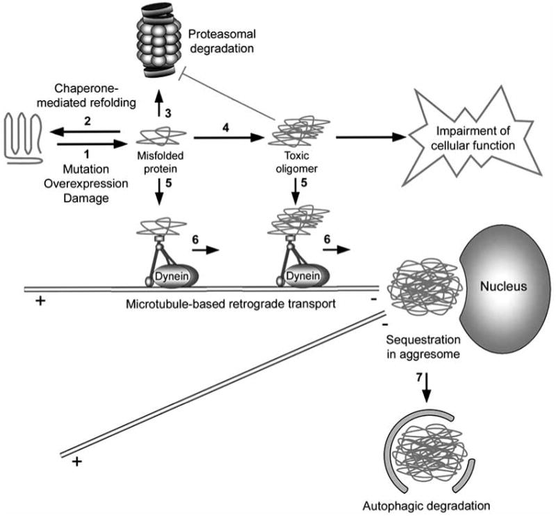
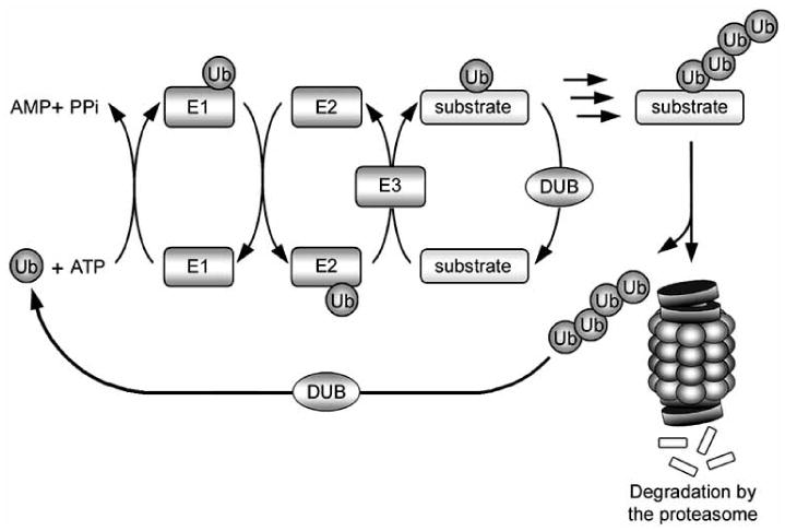
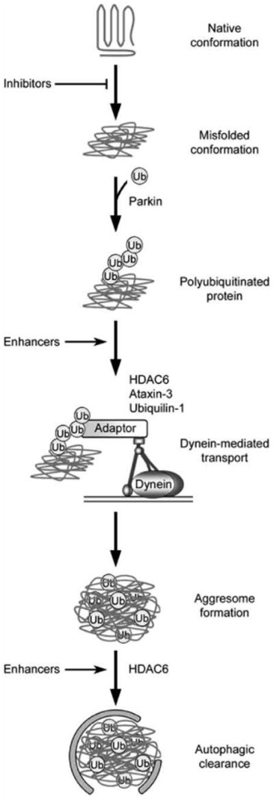
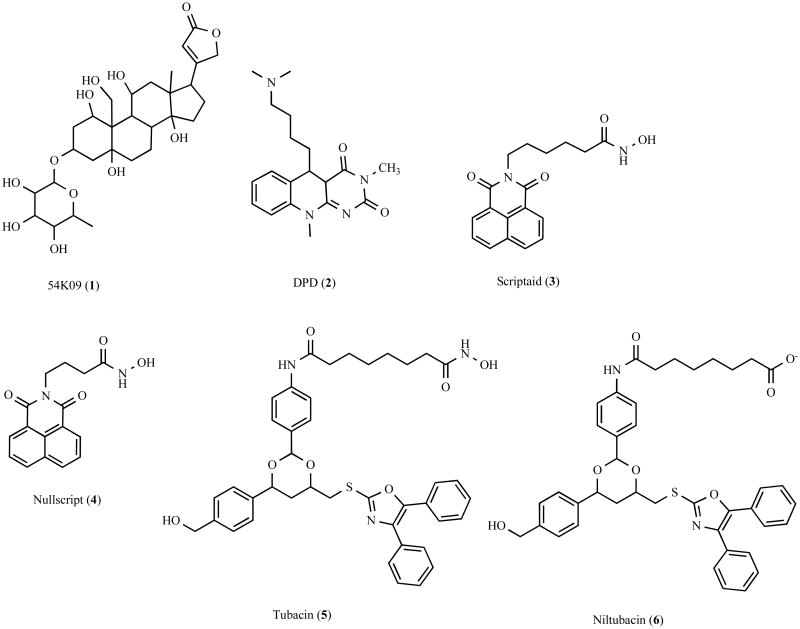
Accumulation of misfolded proteins in proteinaceous inclusions is a prominent pathological feature common to many age-related neurodegenerative diseases, including Parkinson's disease, Alzheimer's disease, Huntington's disease, and amyotrophic lateral sclerosis. In cultured cells, when the production of misfolded proteins exceeds the capacity of the chaperone refolding system and the ubiquitin-proteasome degradation pathway, misfolded proteins are actively transported to a cytoplasmic juxtanuclear structure called an aggresome. Aggresome formation is recognized as a cytoprotective response serving to sequester potentially toxic misfolded proteins and facilitate their clearance by autophagy. Recent evidence indicates that aggresome formation is mediated by dynein/dynactin-mediated microtubule-based transport of misfolded proteins to the centrosome and involves several regulators, including histone deacetylase 6, E3 ubiquitin-protein ligase parkin, deubiquitinating enzyme ataxin-3, and ubiquilin-1. Characterization of the molecular mechanisms underlying aggresome formation and its regulation has begun to provide promising therapeutic targets that may be relevant to neurodegenerative diseases. In this review, we provide an overview of the molecular machinery controlling aggresome formation and discuss potential useful compounds and intervention strategies for preventing or reducing the cytotoxicity of misfolded and aggregated proteins.


Figures

Fig. 1
Fig. 1. Aggresome formation is a cellular defense against the accumulation of aggregated proteins
Genetic mutation, increased protein levels, and oxidative stress can induce protein misfolding (step 1). Once formed, misfolded proteins may be refolded/stabilized by chaperones (step 2) or degraded by the 26S proteasome (step 3). However, when the chaperone and proteasome systems are damaged or overwhelmed, misfolded proteins have the potential to aggregate (step 4) and impair cellular function, such as the inhibition of the proteasomal function. The cell recognizes misfolded and aggregated proteins and transports these proteins to aggresomes in a process mediated by the dynein motor complex (steps 5 and 6). Aggresomes not only sequester potentially harmful aggregated proteins, but also concentrate aggregated proteins for more efficient autophagic degradation (step 7).
Fig. 2
Fig. 2. The ubiquitin-proteasome system
Ubiquitin is covalently attached to a substrate protein through a series of sequential reactions involving three enzymes: an E1 ubiquitin-activating enzyme, which forms a thiol-ester linkage with ubiquitin; an E2 ubiquitin-conjugating enzyme, which transiently carries ubiquitin via a thiol-ester linkage; and finally an E3 ubiquitin-protein ligase, which facilitates the transfer of ubiquitin from the E2 enzyme to the substrate. Successive reactions result in the attachment of a polyubiquitin chain, which targets the substrate for degradation by the 26S proteasome. The polyubiquitin chain is removed from the substrate and recycled by deubiquitinating enzymes (DUBs).
Fig. 3
Fig. 3. Potential steps in the aggresome-autophagy pathway for therapeutic intervention
In this hypothetical model, an initiating event results in the generation of misfolded proteins, which are recognized and polyubiquitinated by an E3 ubiquitin-protein ligase such as parkin. Adaptor proteins, which may include HDAC6, ataxin-3, and ubiquilin-1, link the polyubiquitinated proteins to the dynein motor complex for retrograde transport to the aggresome. Autophagic machinery is recruited to the aggresome in a process involving HDAC6, and aggresomes are degraded. Multiple steps of this pathway could be targeted for treatment of neurodegenerative disease, including the use of small molecules to inhibit protein misfolding, enhance the coupling of misfolded proteins to dynein for retrograde transport, or enhance autophagic clearance of aggresomes. Furthermore, additional intervention strategies could potentially target any step along this pathway.
Fig. 4
Fig. 4
Chemical structure of aggresome inhibitors and their analogues.
Fig. 5
Fig. 5
Chemical structure of aggresome enhancers and their analogues.
Fig. 6
Fig. 6
Chemical structure of bifunctional molecules that promote protein complex formation.
Fig. 7
Fig. 7
Chemical structure of ubistatins, a class of small molecules that selectively bind K48-linked polyubiquitin chains.

References

    1. Ross CA, Poirier MA. Nat Med. 2004;10:S10–17. - PubMed
    1. Chiti F, Dobson CM. Annu Rev Biochem. 2006;75:333–366. - PubMed
    1. Bucciantini M, Giannoni E, Chiti F, Baroni F, Formigli L, Zurdo J, Taddei N, Ramponi G, Dobson CM, Stefani M. Nature. 2002;416:507–511. - PubMed
    1. Kayed R, Head E, Thompson JL, McIntire TM, Milton SC, Cotman CW, Glabe CG. Science. 2003;300:486–489. - PubMed
    1. Fandrich M, Fletcher MA, Dobson CM. Nature. 2001;410:165–166. - PubMed

Publication types

MeSH terms

Substances