. 2008 Apr;9(4):510-27.
doi: 10.1111/j.1600-0854.2008.00710.x. Epub 2008 Jan 22.

Dynactin function in mitotic spindle positioning

Affiliations

Dynactin function in mitotic spindle positioning

Jeffrey K Moore et al. Traffic. 2008 Apr.

Abstract

Dynactin is a multisubunit protein complex necessary for dynein function. Here, we investigated the function of dynactin in budding yeast. Loss of dynactin impaired movement and positioning of the mitotic spindle, similar to loss of dynein. Dynactin subunits required for function included p150(Glued), dynamitin, actin-related protein (Arp) 1 and p24. Arp10 and capping protein were dispensable, even in combination. All dynactin subunits tested localized to dynamic plus ends of cytoplasmic microtubules, to stationary foci on the cell cortex and to spindle pole bodies. The number of molecules of dynactin in those locations was small, less than five. In the absence of dynactin, dynein accumulated at plus ends and did not appear at the cell cortex, consistent with a role for dynactin in offloading dynein from the plus end to the cortex. Dynein at the plus end was necessary for dynactin plus-end targeting. p150(Glued) was the only dynactin subunit sufficient for plus-end targeting. Interactions among the subunits support a molecular model that resembles the current model for brain dynactin in many respects; however, three subunits at the pointed end of brain dynactin appear to be absent from yeast.


Figures

Figure 1
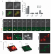
Figure 1. Localization of dynactin in cells
Fluorescent-tagged forms of dynactin subunits are observed at plus ends of microtubules and at SPBs. The α-tubulin subunit Tub1 was tagged with CFP for colocalization in each strain. The width of each image is 5 μm. Strain numbers: yJC3891, Yll049w-3GFP; yJC4147, Nip100-3GFP; yJC5261, Jnm1-3GFP; yJC5389, Arp10-3GFP and yJC5400, Arp1-tdimer2.
Figure 2
Figure 2. Dynamics of dynactin and dynein localization
A) Dynamitin/Jnm1 tagged with tdimer2 and DHC/Dyn1 tagged with 3GFP localized with CFP-labeled microtubules. Dynactin and dynein exhibit a selective association with the daughter-bound set of microtubules. Images were captured on a wide-field microscope. B) Distribution of dynamitin/Jnm1 and DHC on short bipolar spindles in early anaphase cells. From left to right, cells were scored as having fluorescence concentrated on the daughter-bound SPB, concentrated on the mother-bound SPB, on both equally or on neither. The values are the mean of four sets of 50 cells each, and error bars denote standard error of those means. C) Dynein and dynactin colocalize on the sides and ends of cytoplasmic microtubules. Jnm1-tdimer2 and Dyn1-3GFP expressed in num1Δ cells. Three confocal sections at 0.1-μm increments were captured every 10 seconds and collapsed into Z projections using ImageJ. D) Dynactin and dynein colocalize at foci on the cell cortex. Jnm1-tdimer2 and Dyn1-3GFP expressed in wild-type cells expressing CFP-labeled microtubules. Arrowheads denote cortical foci containing both Jnm1 and Dyn1. Images were captured on a wide-field microscope. E) Surface plots of Jnm1-tdimer2 and Dyn1-3GFP images shown in (D) were generated using ImageJ. F) Z projections of 16 confocal sections at 0.2-μm increments in wild-type cells expressing Jnm1-tdimer2 and Dyn1-3GFP. Strains: yJC5652 in panels A,B,D,E and F and yJC5666 in C.
Figure 3
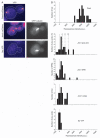
Figure 3. Quantification of dynamitin fluorescence intensity
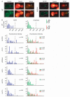
A) Representative images of cells expressing Cse4-mCitrine, Jnm1m-Citrine or CFP-Tub1. Yellow fluorescent protein images were captured using identical settings and pseudocolored with the Fire lookup table in ImageJ. B) Histogram of fluorescence intensity per focus of Cse4-mCitrine in anaphase cells. Thirty-two molecules of Cse4 are present in each spot in late anaphase (28), so the fluorescence intensity per molecule of mCitrine here is 295 arbitrary units (au). C) Histogram of fluorescence intensity of Jnm1-mCitrine in budded cells. Some values are less than zero because of background subtraction. Bud-proximal microtubule plus ends (n = 26) and SPBs (n = 33) were identified using CFP-Tub1. Dashed lines indicate the predicted fluorescence intensities for 4, 8 and 12 molecules of Jnm1-mCitrine. Cortical foci of Jnm1-mCitrine were identified by eye (n = 14). The bottom panel is a control with budded cells expressing CFP-Tub1 and no mCitrine. Here, microtubule plus ends were identified in the CFP channel, and intensity was recorded in the mCitrine channel, n = 12. Strain numbers: Cse4-mCitrine, yJC5463; Jnm1-mCitrine CFP-Tub1, yJC5349 and CFP-Tub1, yJC3883.
Figure 4
Figure 4. Localization of dynactin subunits in null mutants
A diagram illustrates the molecular architecture of vertebrate dynactin, modified with permission from one by Trina Schroer (1). On the bar graphs, the percentage of cells that display localization of a fluorescent-tagged subunit to a given location is plotted for different dynactin null mutants. The fluorescent-tagged subunits are A) p150Glued/Nip100-3GFP, B) dynamitin/Jnm1-mCitrine, C) Yll049w-3GFP, D) Arp10-3GFP and E) Arp1-tdimer2. Strain numbers were as follows: A) Wild-type yJC4147; arp1Δ, yJC4158; jnm1Δ, yJC4156; yll049wΔ, yJC4162 and arp10Δ, yJC5377. B) Wild-type yJC5349; arp1Δ, yJC5381; nip100Δ, yJC5385; yll049wΔ, yJC5387 and arp10Δ, yJC5383. C) Wild-type yJC3891; nip100Δ, yJC4166; arp1Δ, yJC4152; jnm1Δ, yJC4154 and arp10Δ, yJC5379. D) Wild-type yJC5389; arp1Δ, yJC5416; jnm1Δ, yJC5418; nip100Δ, yJC5420 and yll049wΔ,yJC5422.E)Wild-typeyJC5400; nip100Δ/nip100Δ, yJC5483; jnm1Δ/jnm1Δ, yJC5482; yll049wΔ/yll049wΔ, yJC5484 and arp10Δ/arp10Δ, yJC5480. MT, microtubule.
Figure 5
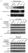
Figure 5. Roles of dynactin subunits in composition of the complex
p150Glued/Nip100-TAP was tagged at the endogenous locus in haploid wild-type and various mutant strains. A) Immunoblots of whole-cell lysates. The level of TAP-tagged p150Glued/Nip100 is similar in wild-type and mutant cells. Dynamitin/Jnm1 is absent from the jnm1 mutant as expected. Cdc3 is a loading control. B) Immunoblots of high-speed supernatants, the S100 fraction, of the whole-cell lysates. The amount of p150Glued/Nip100 is decreased in the jnm1 and yll049w mutants. C) Immunoblots of Nip100-TAP precipitates loaded so that each lane received similar amounts of Nip100. Dynamitin/Jnm1 and p24/Yll049w are important for the association of Arp1 and Arp10 with Nip100. Intensities of Cap2 and Act1 bands were not reproducibly above control (not shown). Arp10 and p24/Yll049w were tagged with 13-myc. Strain numbers: untagged, yJC5813; wild-type, yJC5814; yll04wΔ, yJC5475; jnm1Δ, yJC5817; arp1Δ, yJC5815 and arp10Δ, yJC5816.
Figure 6
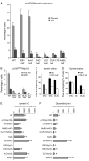
Figure 6. Dynein is important for targeting of p150Glued/Nip100 to the microtubule plus end
A and B) Percentage of cells with Nip100-GFP at a plus end or an SPB in various mutants. DHC, Dyn1. DIC, Pac11. DLIC, Dyn3. DLC, Dyn2. C) Plus-end targeting of GFP-tagged dynein chains in a DLC null mutant, dyn2Δ. D)Plus-end targeting of dynein chains in a DLIC null mutant, dyn3Δ. Dynein heavy chain and DIC require each other for stability and thus for targeting, and no dynein chains target to plus ends in the absence of DIC or intermediate chain. E and F) Fluorescence intensity of Dyn1-3GFP and Jnm1-tdimer2 at the plus end. Values are the means of the fluorescence intensity of the most bud-proximal cytoplasmic microtubule in G2/M cells expressing CFP-Tub1. Error bars denote standard error of the mean. Z-series images were collected for G2/M cells, identified by spindle length and grown in log-phase cultures. Microtubule ends were identified in the CFP-Tub1 image, and intensity measurements were taken from the corresponding plane of the GFP or tdimer2 stack. Asterisks mark results significantly different from wildtype (p < 0.05). Strain numbers and numbers of cells were as follows: A) WT, yJC4147, 369; EB1 null, yJC4177, 124; Num1 null, yJC4316, 301; DHC null, yJC4180, 361; LIS1 null, yJC4178, 210; CLIP-170 null, yJC4182, 124 and NudEL null, yJC4253, 581. B) WT, yJC4147, 369; DHC null, yJC4180, 361; DIC null, yJC5264, 222; DLIC null, yJC4314, 323 and DLC null, yJC43112, 211. C) DHC/WT, yJC2914, 261; DHC/DLC null, yJC4310, 195; DIC/WT, yJC3499, 242; DIC/DLC null, yJC4369, 280; DLIC/WT, yJC3369, 294 and DLIC/DLC null, yJC4367, 270. D) DHC/WT, yJC2914, 261; DHC/DLIC null, yJC4458, 291; DIC/WT, yJC3499, 242; DIC/DLIC null, yJC5266, 235; DLC WT, yJC4555, 166 and DLC/DLIC null, yJC4951, 96. E and F) WT, yJC5652, 23; nip100Δ, yJC5661, 32; pac1Δ, yJC5664, 22; ndl1Δ, yJC5665, 20; bik1Δ, yJC5662, 23; kip2Δ, yJC5663, 13; bim1Δ, yJC5678, 15; dyn1Δ, yJC5669, 16; dyn3Δ, yJC5667, 21; dyn2Δ, yJC5668, 12 and num1Δ, yJC5666, 17.
Figure 7
Figure 7. Dynein accumulation at microtubule plus ends in the absence of dynactin
A) Images of DHC/Dyn1 tagged with 3GFP in wild-type and yll049wΔ cells expressing CFP-labeled microtubules. Arrowheads mark Dyn1-3GFP located at microtubule plus ends. B) Histograms of Dyn1-3GFP fluorescence intensity at microtubule plus ends in wild-type and mutant cells. The fluorescence intensity of Dyn1-3GFP per cytoplasmic microtubule plus end was determined in G2/M and anaphase cells expressing CFP-Tub1-labeled microtubules. Cell cycle stage was assessed from spindle length. Spindles ∼1-1.25 μm in length were scored as G2/M, and longer spindles were scored as anaphase. Blue bars represent microtubule plus ends in G2/M cells. Green bars represent plus ends of cells with properly aligned anaphase spindles that traverse the bud neck. Red bars represent plus ends of cells with anaphase spindles that are contained entirely within the mother cell and misaligned with respect to the axis of division. The wild-type and arp10Δ strains displayed only correctly positioned anaphase spindles. Z-series images were captured from asynchronous cultures of wild-type (yJC4149), nip100Δ (yJC4145), jnm1Δ (yJC4150), yll049wΔ (yJC4160), arp1Δ (yJC4143), arp10Δ (yJC4547) and num1Δ (yJC4164) strains. Microtubule ends were identified using CFP-Tub1, and intensity measurements were taken on the corresponding plane of the GFP stack using ImageJ (Materials and Methods). For G2/M cells, 22-34 microtubule ends were scored per sample, and for anaphase, 21-62 were scored. The p values were generated by t-tests comparing the result for a given mutant with the wild-type result in either G2/M or anaphase. The dashed line on each chart is drawn at the same position for reference.
Figure 8
Figure 8. Model for the function and localization of dyneindynactin with respect to cytoplasmic microtubules and the cell cortex
1) Dynactin localizes to cytoplasmic microtubules through its interaction with dynein. 2) Recruitment of the dynein-dynactin complex to microtubule plus ends. 3) Retention of dynein-dynactin complex at plus ends through interactions with CLIP-170/Bik1, LIS1/Pac1 and NudEL/Ndl1. 4) Dynein and dynactin are offloaded to a cortical-binding site, and dynein is activated to power microtubule sliding.

Similar articles

Cited by

References

    1. Schroer TA. Dynactin. Annu Rev Cell Dev Biol. 2004;20:759–779. - PubMed
    1. Muhua L, Karpova TS, Cooper JA. A yeast actin-related protein homologous to that in vertebrate dynactin complex is important for spindle orientation and nuclear migration. Cell. 1994;78:669–679. - PubMed
    1. Eshel D, Urrestarazu LA, Vissers S, Jauniaux JC, vanVliet-Reedijk JC, Planta RJ, Gibbons IR. Cytoplasmic dynein is required for normal nuclear segregation in yeast. Proc Natl Acad Sci U S A. 1993;90:11172–11176. - PMC - PubMed
    1. Li YY, Yeh E, Hays T, Bloom K. Disruption of mitotic spindle orientation in a yeast dynein mutant. Proc Natl Acad Sci U S A. 1993;90:10096–10100. - PMC - PubMed
    1. Adames NR, Cooper JA. Microtubule interactions with the cell cortex causing nuclear movements in Saccharomyces cerevisiae. J Cell Biol. 2000;149:863–874. - PMC - PubMed

Publication types

MeSH terms