. 2008 Mar;7(2):237-49.
doi: 10.1111/j.1474-9726.2008.00367.x. Epub 2008 Jan 23.

Dystrophin deficiency in Drosophila reduces lifespan and causes a dilated cardiomyopathy phenotype

Affiliations

Dystrophin deficiency in Drosophila reduces lifespan and causes a dilated cardiomyopathy phenotype

Ouarda Taghli-Lamallem et al. Aging Cell. 2008 Mar.

Abstract

A number of studies have been conducted recently on the model organism Drosophila to determine the function of genes involved in human disease, including those implicated in neurological disorders, cancer and metabolic and cardiovascular diseases. The simple structure and physiology of the Drosophila heart tube together with the available genetics provide a suitable in vivo assay system for studying cardiac gene functions. In our study, we focus on analysis of the role of dystrophin (Dys) in heart physiology. As in humans, the Drosophila dys gene encodes multiple isoforms, of which the large isoforms (DLPs) and a truncated form (Dp117) are expressed in the adult heart. Here, we show that the loss of dys function in the heart leads to an age-dependent disruption of the myofibrillar organization within the myocardium as well as to alterations in cardiac performance. dys RNAi-mediated knockdown in the mesoderm also shortens lifespan. Knockdown of all or deletion of the large isoforms increases the heart rate by shortening the diastolic intervals (relaxation phase) of the cardiac cycle. Morphologically, loss of the large DLPs isoforms causes a widening of the cardiac tube and a lower fractional shortening, a phenotype reminiscent of dilated cardiomyopathy. The dilated dys mutant phenotype was reversed by expressing a truncated mammalian form of dys (Dp116). Our results illustrate the utility of Drosophila as a model system to study dilated cardiomyopathy and other muscular-dystrophy-associated phenotypes.


Figures
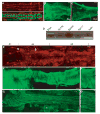
Fig. 1
Fig. 1
Protein distribution in Drosophila embryo and adult heart. (A) Stage 16 wild-type (wt) embryo double-labeled for Dystrophin (Dys, red) and nuclear nmrH15-lacZ (green; see Qian et al., 2005) Dystrophin (Dys) shows dorsal and ventral depositions in the myocardial cells (green nuclei) (B, C, E–G) Adult cardiac tube stained for disc large, (B) Dys, (C) protein outlining the myocardial cell membranes (bar = 25 μm). Note that Dys is localized at the Z-lines in the longitudinal muscles on the side of the myocardial cells. (D) Reverse transcriptase–polymerase chain reaction (RT–PCR) of wt adult heart using specific primers for Dys-like products (DLPs), Dp186, Dp205 and Dp117 compared to control actin (30 PCR cycles). Note that only DLPs and Dp117 are expressed in the adult heart. (E) Dmef2 expression in the adult cardiac tube (A1–A3). Dmef2 is a muscle-specific transcription factor, expressed in all Tinman (arrow, big nuclei) and Seven-up (Svp) expressing myocytes (arrowhead, smaller nuclei and localizes to the ostia) (for further details, see Molina & Cripps, 2001). (F, G) α-Actinin expression in the adult cardiac tube (abdominal segments A1–A3 are shown). (F) Sarcomeric Z-line marker α-actinin shows a spiral or transverse organization of the myofibrils of the contractile myocardium. (F′) High magnification of the myocardium shows the orientation of the myofibrils. (G) α-Actinin reveals a second type of myofibrillar organization in longitudinal non-Tinman-expressing muscles associated with the ventral part of the heart (Molina & Cripps, 2001) (* indicates pericardial cells). (G′) High magnification. For E–G, the bar = 38 μm; for F′, bar = 106 μm; and for G′ bar = 100 μm.
Fig. 2
Fig. 2
Loss of dys function decreases the lifespan in flies. (A) Whole fly reverse transcriptase–polymerase chain reaction indicates the abolishment or reduction in dys isoform transcripts. dysExel6184/dyskx43 flies abolish all but Dp117 transcripts, whereas dysExel6184/dys8-2 and dys8-2/dyskx43 mutants have moderately reduced DLPs transcript levels. (B) Adult expression pattern of GMH5-Gal4 driving with GFP expression specifically in the heart (see also Wessells et al., 2004) (bar = 19 μm). (C) Relative expression of dystrophin in cardiac tube of 1-week-old adults presented as ratio of dys to rp49 mRNA. The bar graph shows that the knockdown of all dys isoforms in dysRNAi/24B-Gal4 flies causes a reduction of ∼60%, and ∼40% with the heart-specific dysRNAi/GMH5-Gal4 knockdown. Each value represents the average ± standard error of the mean of four independent experiments. (D) Survival curves of dys mutant females. The knockdown of all dys isoforms in the mesoderm (dysRNAi/24B-Gal4) shows a moderate reduction in longevity similar to the heterozygous deficiencies dysExel6184/+ and dyskx43/+0. In contrast, the heart-specific dys knockdown does not have an effect on lifespan. Dystrophin-like-product-deficient dysExel6184/dyskx43 mutants show dramatically shortened lifespan. It is possible that this transdeficiency deletes additional genes that are contributing to a normal lifespan.
Fig. 3
Fig. 3
dysExel6184/dyskx43 Mutant flies exhibit age-dependent abnormalities in myofibrillar organization. Representative confocal stacks of adult hearts (posterior A2-anterior A3 segment) stained with α-actinin (green) and anti-Dmef2 (red, right panels) antibodies revealing detail of heart structure. (A-C′) Wild-type (wt) heart structure shows myofibrillar organization in a spiral fashion shown at progressively older different age (A, A′ 1-week-old: 1w; B, B′ 3-week-old: 3w; C, C′ 5-week-old hearts: 5w). The wt hearts at 5w reveal some disruptions of the myocardial myofibrils (C, C′). Ostia at the segmental boundaries of the myocardium were identified as an opening in the heart wall, associated with two pairs of smaller Dmef2-positive nuclei (A′–F′, arrowhead). (D–F′) The dysExel6184/dyskx43 mutant heart structure at 1w (D, D′), 3w (E, E′) and 5w (F, F′), respectively, shows age-dependent disruption of the heart myofibril integrity (D–F arrow pointing out to more spacement between myofibrils). (G) Wild-type and (H) dysExel6184/dyskx43 mutant hearts showing outlined areas devoid of myofibrils in the confocal stacks (H, white circulated area). Areas without myofibrils were measured and normalized to the total area of heart examined (outlined in yellow) using Image J software. See G′ and H′ for a close-up of the myofibril arrangement and that in H′ the asterisk points to a gap between myofibrils (I) Plot of quantification by age of areas devoid of myofibrils (dark areas not stained with α-actinin). Note that dysExel6184/dyskx43 mutant hearts show much more disorganization by this measure than wt. Each data point was from six to eight hearts. P < 0.005 at 1 week; P < 0.0001 at 3 weeks; P < 0.01 at 5 weeks between wt and dysExel6184/dyskx43 mutants. Scale bar = 12 μm.
Fig. 4
Fig. 4
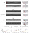
Reduced level or loss of the long dystrophin (Dys)-like product isoforms of Dys results in faster heart rate by shortening the diastolic intervals. (A) Representative M-mode traces (10 s) from high-speed movies of semi-intact flies. Wild-type (wt) flies show rhythmic heart beating at 1-week-old to 3-week-old of age, but moderate arrhythmicity at 5 weeks. dysExel6184/dyskx43 hearts show increased heart rate in 3-week-old and 5-week-old flies. (B) Heart period histograms obtained from 1 min movies plotted as individual data points illustrating the variability of the heart period within a group of flies for controls and dysExel6184/dyskx43 mutants. At 1 week, the mean heart period for wt and dysExel6184/dyskx43 flies is about 0.5 s, which increases in wt to 1 s at 5 weeks. In contrast, aging dysExel6184/dyskx43 shows less of an increase, to only 0.8 s at 5 weeks (bottom panel in B). (C) Standard deviation of the heart period was used as a measure of irregularity in heart rhythm (‘arrhythmicity index’). All time points (except 1 week) show less arrhythmicity for dys mutants compared to wt flies. Differences were estimated by t-test P < 0.05 and are indicated by *. (D) Mean diastolic interval (± SEM) for wt and dys mutant flies obtained from 1 min high-speed movies at the indicated ages. Significant differences were determined by two-tailed independent samples t-test. P values < 0.05 were considered significant (*P < 0.01). (E) Percentage of total flies showing asystoles (prolonged diastolic phases of more than 1 s) in 1-min movie clips. Differences were estimated by t-test P < 0.05 and are indicated by *.
Fig. 5
Fig. 5
Reduced or no dystrophin-like products (DLPs) causes dilated cardiomyopathy in the adult Drosophila heart. (A) Two-segment image of a 1-week-old wild-type (wt) heart in systole (top) and diastole (bottom). (B) 1-week-old dysExel6184/dyskx43 heart in systole and diastole. Note that both diastolic and systolic diameters are wider in the DLPs-deficient mutants compared to wt. Arrowheads indicate the heart wall in both phases of the cardiac cycle. (C–E) Heart parameters in wt and dys mutants. (C,D) dysExel6184/dyskx43 and dys8-2/dyskx43 mutants have a larger systolic (C) and diastolic diameters (D) than the wt at all ages. Significant differences were determined by t-test. P values < 0.05 were considered significant (*P < 0.0001). (E) Plot of fractional shortening for the wt and dys mutants. dysExel6184/dyskx43 shows lower fractional shortening at all ages, whereas dys8-2/dyskx43 mutants only at 3 weeks or older (*P < 0.001, except dys8-2/dyskx43 at 7 weeks is P < 0.05). Movies were taken from 15 to 20 flies for each genotype. (F, G) Rescue of dysExel6184/dyskx43 mutants by the mammalian short dys isoform Dp116. Mesodermal expression of Dp116 restores the dilated systolic diameter (F) and the reduced fractional shortening to near normal (G). The Dp116 transgene was driven by the mesodermal driver 24B-GAL4 in the dys mutant background (Dp116/+; dyskx43/dysExel6184,24B). Cardiac function evaluated in 1-week-old rescue dys mutants shows a normalization of the systolic dysfunction (F, G) (*P < 0.01 by t-test assuming equal variances). For the genotype Dp116/+; dyskx43/dysExel6184,24B measurements (± standard error) were derived from movies of 20 flies. Similar rescue as with 1-week-old flies was observed in 3-week-old flies (data not shown).

Similar articles

Cited by

References

    1. Adams MD, Celniker SE, Holt RA, Evans CA, Gocayne JD, Amanatides PG, Scherer SE, Li PW, Hoskins RA, Galle RF, George RA, et al. The genome sequence of Drosophila melanogaster. Science. 2000;5461:2185–2195. - PubMed
    1. Bassett D, Currie PD. Identification of a zebrafish model of muscular dystrophy. Clin Exp Pharmacol Physiol. 2004;8:537–540. - PubMed
    1. Bessou C, Giugia JB, Franks CJ, Holden-Dye L, Segalat L. Mutations in the Caenorhabditis elegans dystrophin-like gene dys-1 lead to hyperactivity and suggest a link with cholinergic transmission. Neurogenetics. 1998;2:61–72. - PubMed
    1. Bier E. Drosophila, the golden bug, emerges as a tool for human genetics. Nat Rev Genet. 2005;1:9–23. - PubMed
    1. Bier E, Bodmer R. Drosophila, an emerging model for cardiac disease. Gene. 2004;342:1–11. - PubMed

Publication types

MeSH terms

LinkOut - more resources