The pathways connecting the hippocampal formation, the thalamic reuniens nucleus and the thalamic reticular nucleus in the rat
- PMID: 18221482
- PMCID: PMC2408996
- DOI: 10.1111/j.1469-7580.2008.00858.x
The pathways connecting the hippocampal formation, the thalamic reuniens nucleus and the thalamic reticular nucleus in the rat
Abstract
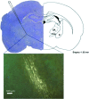
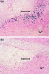
Most dorsal thalamic nuclei send axons to specific areas of the neocortex and to specific sectors of the thalamic reticular nucleus; the neocortex then sends reciprocal connections back to the same thalamic nucleus, directly as well indirectly through a relay in the thalamic reticular nucleus. This can be regarded as a 'canonical' circuit of the sensory thalamus. For the pathways that link the thalamus and the hippocampal formation, only a few comparable connections have been described. The reuniens nucleus of the thalamus sends some of its major cortical efferents to the hippocampal formation. The present study shows that cells of the hippocampal formation as well as cells in the reuniens nucleus are retrogradely labelled following injections of horseradish peroxidase or fluoro-gold into the rostral part of the thalamic reticular nucleus in the rat. Within the hippocampal formation, labelled neurons were localized in the subiculum, predominantly on the ipsilateral side, with fewer neurons labelled contralaterally. Labelled neurons were seen in the hippocampal formation and nucleus reuniens only after injections made in the rostral thalamic reticular nucleus (1.6-1.8 mm caudal to bregma). In addition, the present study confirmed the presence of afferent connections to the rostral thalamic reticular nucleus from cortical (cingulate, orbital and infralimbic, retrosplenial and frontal), midline thalamic (paraventricular, anteromedial, centromedial and mediodorsal thalamic nuclei) and brainstem structures (substantia nigra pars reticularis, ventral tegmental area, periaqueductal grey, superior vestibular and pontine reticular nuclei). These results demonstrate a potential for the thalamo-hippocampal circuitry to influence the functional roles of the thalamic reticular nucleus, and show that thalamo-hippocampal connections resemble the circuitry that links the sensory thalamus and neocortex.
Figures


References
-
- Abercrombie M. Estimation of nuclear population from microtome sections. Anat Rec. 1946;94:239–247. - PubMed
-
- Aggleton JP, Desimone R, Mishkin M. The origin, course, and termination of the hippocampothalamic projections in the macaque. J Comp Neurol. 1986;243:409–421. - PubMed
-
- Aker RG, Ozyurt HB, Yananli HR, et al. GABA(A) Y receptor mediated transmission in the thalamic reticular nucleus of rats with genetic absence epilepsy shows regional differences: functional implications. Brain Res. 2006a;21:213–221. - PubMed
-
- Aker RG, Yananlı HR, Gurbanova AA, et al. Amygdala kindling in the WAG/Rij rat model of absence epilepsy. Epilepsia. 2006b;47:33–40. - PubMed
-
- Avanzini G, de Curtis M, Marescaux C, Panzica F, Spreafico R, Vergnes M. Role of the thalamic reticular nucleus in the generation of rhythmic thalamo-cortical activities subserving spikes and waves. J Neural Transm. 1992;35:85–95. - PubMed
Publication types
MeSH terms
Substances
LinkOut - more resources
Full Text Sources
Miscellaneous

