Comparative Study
. 2008 Jan 25:9:14.
doi: 10.1186/1471-2202-9-14.

Discordant localization of WFA reactivity and brevican/ADAMTS-derived fragment in rodent brain

Affiliations
Comparative Study

Discordant localization of WFA reactivity and brevican/ADAMTS-derived fragment in rodent brain

Joanne M Ajmo et al. BMC Neurosci. .

Abstract

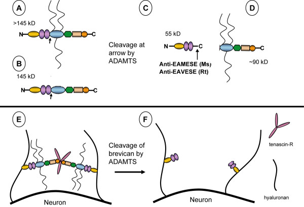
Background: Proteoglycan (PG) in the extracellular matrix (ECM) of the central nervous system (CNS) may act as a barrier for neurite elongation in a growth tract, and regulate other characteristics collectively defined as structural neural plasticity. Proteolytic cleavage of PGs appears to alter the environment to one favoring plasticity and growth. Brevican belongs to the lectican family of aggregating, chondroitin sulfate (CS)-bearing PGs, and it modulates neurite outgrowth and synaptogenesis. Several ADAMTSs (a disintegrin and metalloproteinase with thrombospondin motifs) are glutamyl-endopeptidases that proteolytically cleave brevican. The purpose of this study was to localize regions of adult CNS that contain a proteolytic-derived fragment of brevican which bears the ADAMTS-cleaved neoepitope sequence. These regions were compared to areas of Wisteria floribunda agglutin (WFA) reactivity, a common reagent used to detect "perineuronal nets" (PNNs) of intact matrix and a marker which is thought to label regions of relative neural stability.

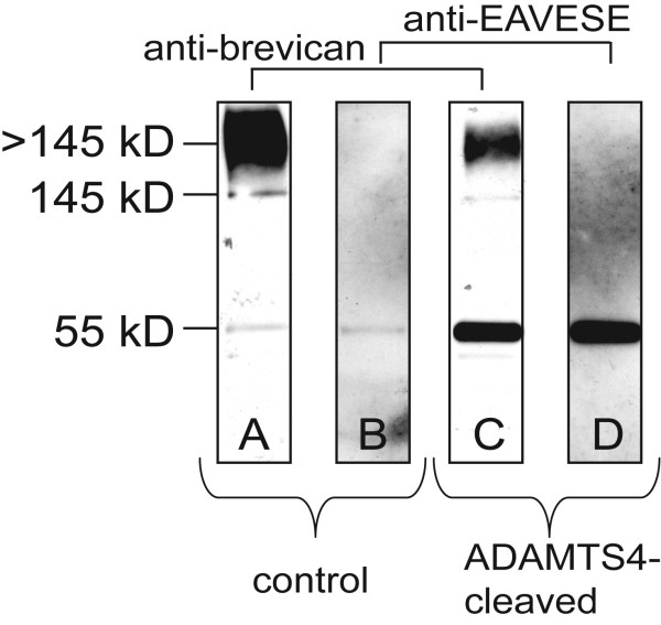
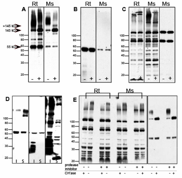
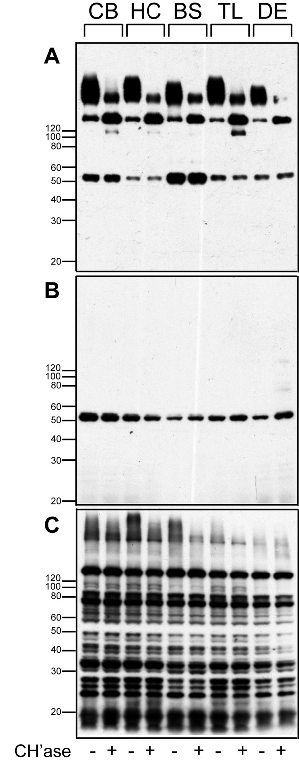
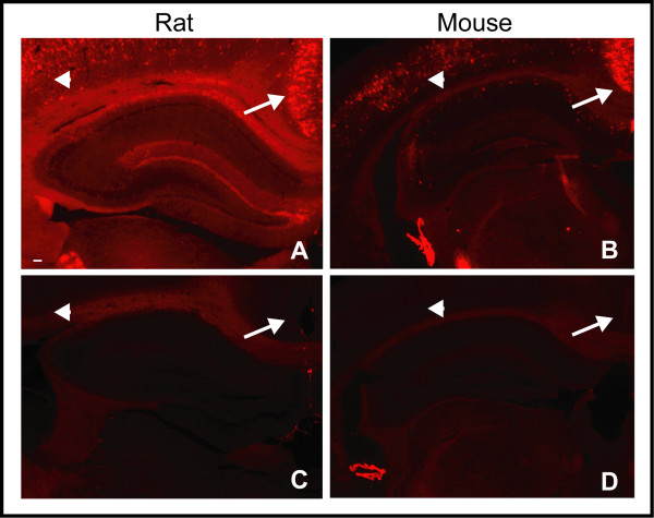
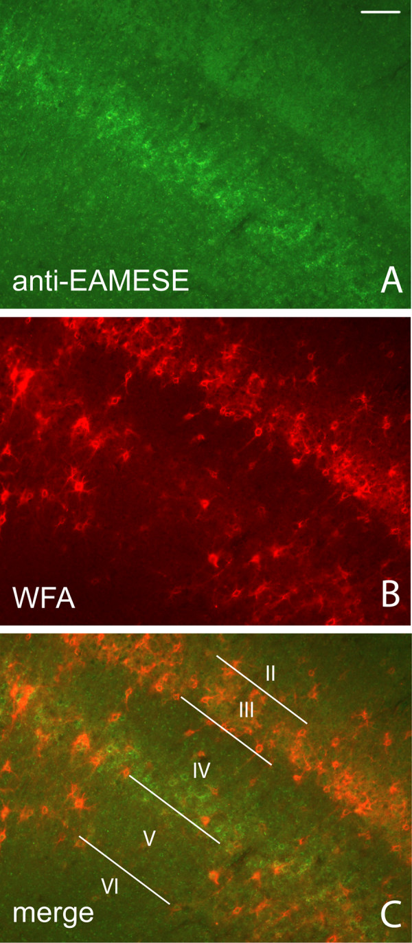
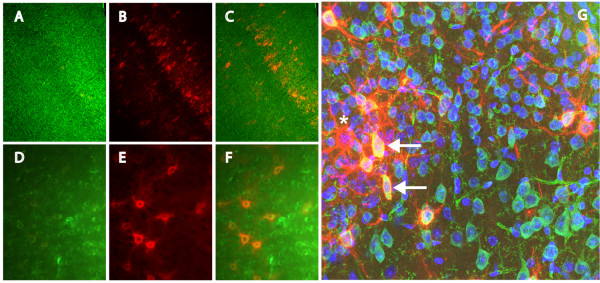
Results: WFA reactivity was found primarily as PNNs, whereas brevican and the ADAMTS-cleaved fragment of brevican were more broadly distributed in neuropil, and in particular regions localized to PNNs. One example is hippocampus where the ADAMTS-cleaved brevican fragment is found surrounding pyramidal neurons, in neuropil of stratum oriens/radiatum and the lacunosum moleculare. The fragment was less abundant in the molecular layer of the dentate gyrus. Mostly PNNs of scattered interneurons along the pyramidal layer were identified by WFA. In lateral thalamus, the reticular thalamic nucleus stained abundantly with WFA whereas ventral posterior nuclei were markedly immunopositive for ADAMTS-cleaved brevican. Using Western blotting techniques, no common species were reactive for brevican and WFA.

Conclusion: In general, a marked discordance was observed in the regional localization between WFA and brevican or the ADAMTS-derived N-terminal fragment of brevican. Functionally, this difference may correspond to regions with varied prevalence for neural stability/plasticity.


Figures

Figure 1
Figure 1
Brevican processing and fragments formed by ADAMTS cleavage. Schematic representation of brevican, its endogenous proteolytic fragments and their interaction with other components of brain ECM: (A) Secreted brevican core protein which bears 1–3 chondroitin sulfate chains (MW > 145 kD). (B) Secreted brevican core protein without chondroitin sulfate chains (MW = 145 kD). When cleaved by extracellular glutamyl-endopeptidases, the ADAMTSs (arrows in A and B), an N-terminal, 55 kD fragment is formed (C) that contains a unique C-terminal murine (Ms) epitope sequence "EAMESE", homologous to the rat (Rt) "EAVESE" which are selectively recognized by respective neoepitope antibodies, anti-EAMESE or anti-EAVESE (C). A larger C-terminal fragment is formed upon ADAMTS cleavage (D). The > 145 kD and 145 kD isoforms of brevican or other lecticans in matrix form an tertiary aggregate complex with hyaluronan and tenascin-R (E) and when cleaved by ADAMTSs, the proteolytic degradation of brevican "loosens" the ECM complex (F).
Figure 2
Figure 2
N-terminal and neoepitope fragment formed by ADAMTS cleavage of brevican. Recombinant ADAMTS4 cleavage product of rat brevican is recognized by anti-EAVESE: (A) Proteoglycan-containing fraction eluates of rat brain extracts from a DEAE cation exchange matrix were probed with anti-brevican. (B) Proteoglycan fraction was probed with anti-EAVESE, antibody raised against the C-terminal neoepitope sequence of the ADAMTS-cleaved N-terminal fragment of brevican. (C) Proteoglycan fraction after incubation with recombinant ADAMTS4 and probed with anti-brevican or (D) anti-EAVESE. After incubation with human recombinant ADAMTS4, brevican was proteolytically cleaved resulting in diminished full length brevican and the appearance of a 55 kD N-terminal fragment recognized by both anti-brevican and anti-EAVESE.
Figure 3
Figure 3
Comparison of brevican, the ADAMTS-derived neoepitope of brevican and WFA reactivity in rat and mouse brain extracts. Western blot of brevican, EAV(M)ESE, and Wisteria floribunda agglutinin (WFA) in rodent brain extracts before and after chondroitinase digestion: Rat (Rt) and mouse (Ms) extracts were probed for (A) anti-brevican, (B) anti-EAVESE (Rt) or anti-EAMESE (Ms), or (C) biotinylated WFA: Samples were treated with (+) and without (-) Chondroitinase ABC (Chase). (A+) The 145 kD core protein of brevican increased after Chase treatment, (B+) the proteolytic brevican fragment remained unchanged, and (C+) only a high molecular weight WFA-reactive band was diminished in Ms. (C) After probing with WFA, multiple, unidentified lower molecular weight bands were observed along with less abundant, high molecular weight moieties. The right panel in (C) was probed with secondary, HRP-conjugated streptavidin alone, which revealed two, major non-specific bands. (D) After differential centrifugation of rat brain tissue, brevican immunoreactivity (left panel) was predominately found in the soluble fraction (S), whereas most of the WFA reactivity (right panel) was observed in the membrane "insoluble" fraction (I) whereas anti-EAVESE immunoreactivity (middle panel) was evident in both fractions. (E) Rt and Ms samples were treated with Chase in the absence (-) and presence (+) of a protease inhibitor cocktail (left panel) and probed with biotinylated-WFA. The high molecular weight smear is eliminated after treatment with Chase, but the protease inhibitor did not change the pattern. (right panel) The same membrane was probed with anti-brevican where complete removal of CS chains led to an increase in abundance of the core protein with no change in the abundance of fragment. Protease inhibitor had no effect on Chase action.
Figure 4
Figure 4
Regional distribution of brevican, EAMESE and WFA reactivity in rat brain. Western blot of brevican, EAMESE, and biotinylated-Wisteria floribunda agglutinin (WFA) in extracts from various regions of mouse brain before and after Chase digestion: Cerebellum (CB), hippocampus (HC), brain stem (BS), temporal lobe (TL) and diencephalon (DE)) extracts were probed for (A) anti-brevican, (B) anti-EAMESE and (C) biotinylated-WFA. Samples were pre-treated with (+) and without (-) Chase. (A+) The 145 kD core protein of mouse brevican increased after chondroitinase treatment, (B+) the proteolytic fragment remained unchanged, and (C+) high molecular weight, biotinylated WFA reactive smears were only slightly affected by Chase.
Figure 5
Figure 5
Chondroitinase treatment of brain sections prior to binding by WFA. Binding of Wisteria floribunda agglutinin (WFA) lectin after treatment with Chase. Paraformaldehyde-fixed coronal sections from (A and C) rat and (B and D) mouse brain were probed with biotinylated-WFA (A and B) before and (C and D) after Chase treatment. Arrow = retrosplenial cortex; arrow head = parietal cortex. Note the near elimination of reactivity after Chase treatment. All images were captured at 25× magnification. Scale bar represents 100 μm.
Figure 6
Figure 6
Histochemical localization of WFA and ADAMTS-derived brevican fragment in rat and mouse brain. Localization of Wisteria floribunda agglutinin (WFA) and ADAMTS-derived fragment of brevican in rat and mouse brain including lateral thalamus (A-F), hippocampus (G-L) and cerebellum (M-R): Epifluorescent micrographs of biotinylated-WFA reactivity (A, D, G, J, M, and P, red), anti-EAVESE (rat) immunoreactivity (B, H, N, green), anti-EAMESE (mouse) immunoreactivity (E, K, Q, green) and merged composites of WFA and anti-EAVESE (C, I O), and WFA and anti-EAMESE (F, L, R) in fixed brain sections. Images A-L were captured at 25× magnification and M-R were captured at 100× magnification. Scale bar represents 100 μm.
Figure 7
Figure 7
Identification of PNNs with antibody against the ADAMTS-derived fragment of brevican. Mouse PNNs immunoreactive for the ADAMTS-derived fragment of brevican distinguished with anti-EAMESE (green) (A) were broadly distributed in cerebral cortex, but were especially prominent deep in cortical layer IV, which differs from the distinct pattern of WFA staining of PNNs (B). The most intense region of WFA reactivity appears in cortical layer III of primary somatosensory cortex (B-C). Scattered WFA positive PNNs are also found in layer V. Images A-C were captured at 100× magnification. Scale bar represents 100 μm.
Figure 8
Figure 8
Localization of WFA and brevican reactivity in PNNs. Brevican immunoreactivity was found in (A and C) neuropil and PNNs of cortical layers II, III, deep layer IV and V. WFA immunoreactivity is predominant in (B and C) PNNs of cortical layer III. A higher magnification of cortex reveals PNNs that are positive for (D) brevican and (E) WFA reactivity. A confocal micrograph (G) of retrosplenial cortex stained with anti-brevican (green), biotinylated-WFA (red) and DAPI (blue) demonstrates a small subpopulation reactive for both anti-brevican and WFA (arrows). While the majority of PNNs showed only brevican immunoreactivity, some were identified as only WFA reactive (denoted by asterisk). Images A-C were captured at 100× magnification, D-F were captured at 200× magnification and confocal image G was captured at 630×. Scale bar represents 100 μm.

References

    1. Herndon ME, Lander AD. A diverse set of developmentally regulated proteoglycans is expressed in the rat central nervous system. Neuron. 1990;4:949–961. doi: 10.1016/0896-6273(90)90148-9. - DOI - PubMed
    1. Lundell A, Olin AI, Morgelin M, al-Karadaghi S, Aspberg A, Logan DT. Structural basis for interactions between tenascins and lectican C-type lectin domains: evidence for a crosslinking role for tenascins. Structure. 2004;12:1495–1506. doi: 10.1016/j.str.2004.05.021. - DOI - PubMed
    1. Yamaguchi Y. Lecticans: organizers of brain extracellular matrix. Cell Mol Life Sci. 2000;57:276–289. doi: 10.1007/PL00000690. - DOI - PMC - PubMed
    1. Young NM, Williams RE. Assignment of lectins specific for D-galactose or N-acetyl-D-galactosamine to two groups, based on their circular dichroism. Can J Biochem Cell Biol. 1985;63:268–271. - PubMed
    1. Brauer K, Hartig W, Bigl V, Bruckner G. Distribution of parvalbumin-containing neurons and lectin-binding perineuronal nets in the rat basal forebrain. Brain Res. 1993;631:167–170. doi: 10.1016/0006-8993(93)91205-7. - DOI - PubMed

Publication types

MeSH terms

LinkOut - more resources