Review
. 2008 Aug;1779(8):495-506.
doi: 10.1016/j.bbagrm.2007.12.010. Epub 2008 Jan 5.

Roles of ubiquitination at the synapse

Affiliations
Review

Roles of ubiquitination at the synapse

Kevin F Haas et al. Biochim Biophys Acta. 2008 Aug.

Abstract

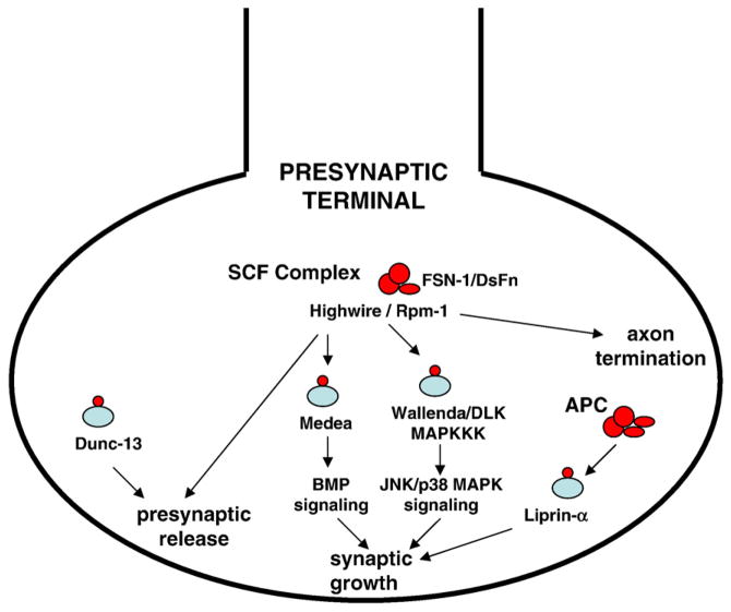
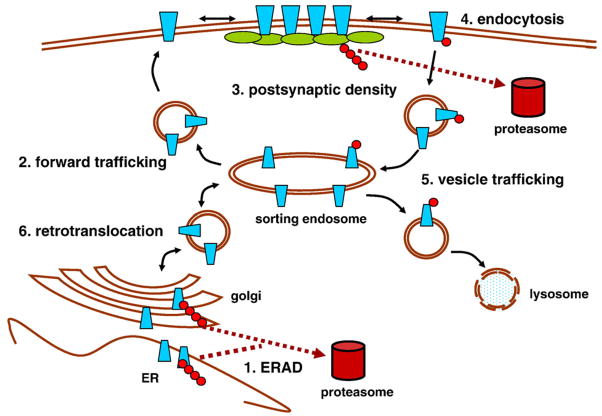
The ubiquitin proteasome system (UPS) was first described as a mechanism for protein degradation more than three decades ago, but the critical roles of the UPS in regulating neuronal synapses have only recently begun to be revealed. Targeted ubiquitination of synaptic proteins affects multiple facets of the synapse throughout its life cycle; from synaptogenesis and synapse elimination to activity-dependent synaptic plasticity and remodeling. The recent identification of specific UPS molecular pathways that act locally at the synapse illustrates the exquisite specificity of ubiquitination in regulating synaptic protein trafficking and degradation events. Synaptic activity has also been shown to determine the subcellular distribution and composition of the proteasome, providing additional mechanisms for locally regulating synaptic protein degradation. Together these advances reveal that tight control of protein turnover plays a conserved, central role in establishing and modulating synapses in neural circuits.


Figures

Fig. 1
Fig. 1
UPS presynaptic pathways. UPS pathways regulate presynaptic differentiation and neurotransmission strength. In Drosophila and C. elegans, the large E3 ligase Highwire/Rpm-1 operates as part of a unique SCF complex that includes the F-box protein FSN-1/DsFn, which targets the MAPKKK Wallenda/DLK and regulates synaptic growth via JNK MAPK or p38 MAPK signaling, respectively. In C. elegans, Rpm-1 regulates axon termination through a separate incompletely characterized pathway. In Drosophila, Highwire also regulates presynaptic neurotransmitter release, as does targeted ubiquitination of dUNC-13, a synaptic vesicle priming protein. The APC E3 ligase complex operates presynaptically to regulate synaptic growth by targeting Liprin-α.
Fig. 2
Fig. 2
UPS postsynaptic mechanisms regulating neurotransmitter receptors. There are multiple modes of UPS regulation of receptors in the postsynaptic density. 1. ERAD: Receptor subunits are targeted for degradation by the proteasome after ubiquitination and translocation from the ER. 2. Forward trafficking: Ubiquitination regulates the rate and efficiency of forward trafficking from the ER to the plasma membrane. 3. Scaffold control: Polyubiquitination and targeted degradation of postsynaptic density scaffolding proteins determines the number of receptor slots. 4. Membrane recycling: Mono- or polyubiquitination of receptors and/or endocytic machinery triggers endocytosis. 5. Vesicle trafficking: Monoubiquitination serves as a trafficking signal, directing receptors to the late endosome for lysosomal degradation. 6. Retrotranslocation: Reverse trafficking of surface membrane receptors back to the ER can lead to extracellular domain polyubiquitination and translocation from the membrane for proteasome degradation.
Fig. 3
Fig. 3
Genetically targeted postsynaptic proteasome inhibition acutely regulates class-specific glutamate receptor expression. Drosophila genetic targeting of postsynaptic proteasome inhibition was induced by feeding third instar larvae RU486-containing food for a brief interval (6 h). Confocal imaging of a NMJ synapse was performed in control (−RU486) and proteasome-blocked (+RU486) animals. A, Representative images from synapses co-labeled for A-class (GluRIIA) or B-class (GluRIIB) receptors and synaptic membrane marker (HRP). Smaller images show higher magnification of individual synaptic boutons labeled for GluRIIA or GluRIIB. B, GluR expression intensity was quantified for GluRIIA and GluRIIB relative to HRP. GluRIIA was decreased and GluRIIB was increased by proteasome blockade. Bars represent mean± SEM, *p<0.05, ***p<0.001.

References

    1. Etlinger JD, Goldberg AL. A soluble ATP-Dependent proteolytic system responsible for the degradation of abnormal proteins in reticulocytes. PNAS. 1977;74:54–58. - PMC - PubMed
    1. Hershko A, Ciechanover A, Heller H, Haas AL, Rose IA. Proposed role of ATP in protein breakdown — conjugation of proteins with multiple chains of the polypeptide of ATP-Dependent proteolysis. PNAS. 1980;77:1783–1786. - PMC - PubMed
    1. Jiang YH, Beaudet AL. Human disorders of ubiquitination and proteasomal degradation. Curr Opin Pediatr. 2004;16:419–426. - PubMed
    1. Reinstein E, Clechanover A. Protein degradation and human diseases: the ubiquitin connection. Ann Intern Med. 2006;145:676–684. - PubMed
    1. Ciechanover A, Brundin P. The ubiquitin proteasome system in neurodegenerative diseases: sometimes the chicken, sometimes the egg. Neuron. 2003;40:427–446. - PubMed

Publication types

Substances

LinkOut - more resources