. 2008 Jun 1;63(11):1013-21.
doi: 10.1016/j.biopsych.2007.11.021. Epub 2008 Jan 28.

Distinct roles of adenylyl cyclases 1 and 8 in opiate dependence: behavioral, electrophysiological, and molecular studies

Affiliations

Distinct roles of adenylyl cyclases 1 and 8 in opiate dependence: behavioral, electrophysiological, and molecular studies

Venetia Zachariou et al. Biol Psychiatry. .

Abstract

Background: Opiate dependence is a result of adaptive changes in signal transduction networks in several brain regions. Noradrenergic neurons of the locus coeruleus (LC) have provided a useful model system in which to understand the molecular basis of these adaptive changes. One of most robust signaling adaptations to repeated morphine exposure in this brain region is upregulation of adenylyl cyclase (AC) activity. Earlier work revealed the selective induction of two calmodulin-dependent AC isoforms, AC1 and AC8, after chronic morphine, but their role in opiate dependence has remained unknown.

Methods: Whole cell recordings from LC slices, behavioral paradigms for dependence, and gene array technology have been used to dissect the role of AC1 and AC8 in chronic morphine responses.

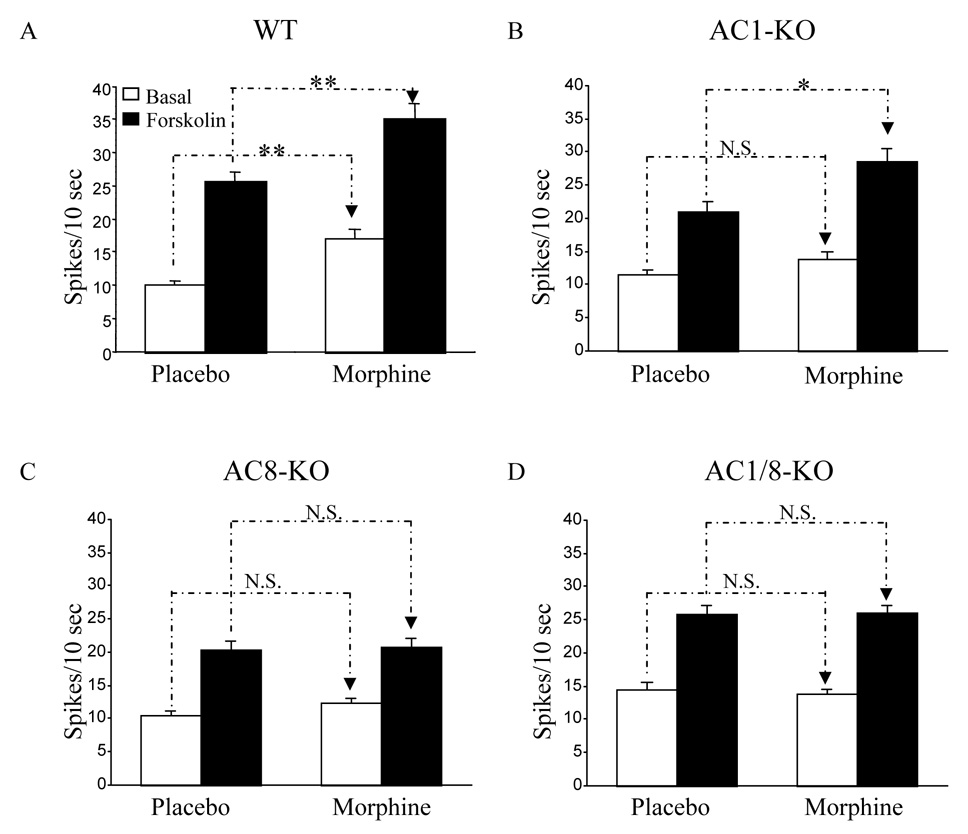
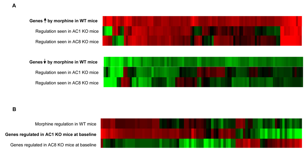
Results: Both AC1 and AC8 knockout mice exhibit reduced opiate dependence on the basis of attenuated withdrawal; however, partially distinct withdrawal symptoms were affected in the two lines. Loss of AC1 or AC8 also attenuated the electrophysiological effects of morphine on LC neurons: knockout of either cyclase attenuated the chronic morphine-induced enhancement of baseline firing rates as well as of regulation of neuronal firing by forskolin (an activator of ACs). The DNA microarray analysis revealed that both AC1 and AC8 affect gene regulation in the LC by chronic morphine and, in addition to common genes, each cyclase influences the expression of a distinct subset of genes.

Conclusions: Together, these findings provide fundamentally new insight into the molecular and cellular basis of opiate dependence.


Conflict of interest statement

Disclosure of Biomedical Financial Interests and potential conflicts of interest: Dr. Venetia Zachariou, Dr. Rongjian Liu, Mr. Quincey Laplant, Dr. Guanghua Xiao, Mr. William Renthal, Dr. Guy C. Chan, Dr. Daniel R. Storm, Dr. George Aghajanian and Dr. Eric J. Nestler report no financial Interests and potential conflicts of interest.

Figures

Figure 1
Figure 1. Knockout of AC1 decreases morphine dependence
A. AC1 KO mice and wildtype littermate control mice, treated chronically with morphine, were injected with naloxone (1 mg/kg, s.c.) and opiate withdrawal signs were monitored for 25 min (n=5 per genotype). B. A different group of animals was injected with morphine once a day (20 mg/kg) and analgesic responses were monitored in a 52°C hot plate test for four consecutive days (n=8–10 per genotype). Responses are expressed as % maximal possible effect (see Materials and Methods). Data are expressed as means ± S.E.M., *p<0.05 for genotype over treatment, two-way ANOVA followed by Bonferroni test.
Figure 2
Figure 2. Knockout of AC8 decreases morphine dependence
AC8 KO mice and wildtype littermate control mice were treated as described in the legend to Fig. 1. Data are expressed as means ± S.E.M., *p<0.05 for genotype over treatment, two-way ANOVA followed by Bonferroni test.
Figure 3
Figure 3. Double knockout of AC1 and AC8 decreases morphine dependence
AC1/8 double KO mice and wildtype littermate control mice were treated as described in the legend to Fig. 1. Data are expressed as means ± S.E.M., *p<0.05 for genotype over treatment, two-way ANOVA followed by Bonferroni test.
Figure 4
Figure 4. Enhanced baseline and forskolin-induced firing of LC neurons after chronic morphine treatment was attenuated in AC1, AC8, and AC1/8 KO mice
Bar graphs summarize data for LC firing rates in brain slices from placebo- and morphine-treated mice at baseline and in response to forskolin (10 µM, 10–15 min). These parameters were studied in WT mice (n=24 mice, 67 cells) (A), AC1 KO mice (n=10 mice, 34 cells) (B), AC8 KO mice (n=10 mice, 38 cells) (C), and AC1/8 double KO mice (n=11 mice, 42 cells) (D). Note that data from wildtype mice of each mutant line were combined since no differences were observed among them. As shown in the figure there is a blunting of the effect of chronic morphine on baseline firing rates in all three lines of KO and a complete block of the enhanced effect of forskolin on firing rates after chronic morphine in AC8 and AC1/8 KO’s, with a partial blunting effect in the AC 1 KO. Data shown as mean ± SEM; *p < 0.05 ; **P < 0.01 (One-way ANOVA followed by Turkey test)
Figure 5
Figure 5. Morphine regulation of DAMGO sensitivity in LC neurons of AC1, AC8 and AC1/8 double KO mice
Concentration-response curves for the µ-opioid receptor agonist DAMGO were measured as percentage suppression of baseline firing rate after stepwise increases in DAMGO concentration in perfusate. Four groups each of non-dependent (placebo-treated) and dependent (morphine-treated) mice were tested: wildtype (WT) (A), AC1 KO (B), AC8 KO (C), and AC1/8 double KO’s (D). As expected from previous studies, WT mice in all three KO lines showed a rightward shift in the concentration response curve (decreased sensitivity), hence the data were collapsed into a single WT group. In contrast to WT, all three AC KO groups showed little or no rightward shift after morphine treatment, but showed decreased sensitivity to DAMGO relative to WT in the non-dependent condition (see Table for IC50 comparisons).
Figure 6
Figure 6. Morphine regulation of gene expression in LC of AC1 and AC8 KO mice
A. The upper row of the two heatmaps illustrate that genes that are significantly upregulated (red) or downregulated (green) in the LC of wildtype (WT) mice after chronic morphine administration. The two lower rows of each heatmap illustrate how those same genes were affected by chronic morphine in AC1 or AC8 KO mice. Note that while many genes are similarly regulated by morphine in WT and KO mice, roughly half of the genes normally regulated by morphine are no longer altered in each respective KO. Interestingly, certain genes are even oppositely regulated uniquely in AC1 or AC8 KO mice. B. Middle row in heatmap illustrates genes that are significantly altered in LC of sham-treated AC1 KO mice compared to WT littermate controls. The other rows illustrate how those same genes are affected by chronic morphine in WT mice (upper row) and in sham-treated AC8 KO mice compared to WT littermate controls. Note how loss of AC1 at baseline induces a regulatory pattern reminiscent of the effects of morphine in WT mice, an effect not seen in AC8 KO mice.

Comment in

References

    1. Aghajanian GK. Tolerance of locus coeruleus neurons to morphine and suppression of withdrawal response by clonidine. Nature. 1978;276:186–188. - PubMed
    1. Taylor JR, Elsworth JD, Garcia EJ, Grant SJ, Roth RH, Redmond DE., Jr Clonidine infusions into the locus coeruleus attenuate behavioral and neurochemical changes associated with naloxone-precipitated withdrawal. Psychopharmacology. 1988;96:121–134. - PubMed
    1. Koob GF, Maldonado R, Stinus L. Neural substrates of opiate withdrawal. Trends Neurosci. 1992;15:186–191. - PubMed
    1. Maldonado R, Koob GF. Destruction of the locus coeruleus decreases physical signs of opiate withdrawal. Brain Res. 1993;605:128–138. - PubMed
    1. Aston-Jones G, Hirata A, Akaoka H. Local opiate withdrawal in locus coeruleus in vivo. Brain Res. 1997;765:331–336. - PubMed

MeSH terms