. 2008 Jan;20(1):186-200.
doi: 10.1105/tpc.107.055178. Epub 2008 Jan 25.

EST analysis of hop glandular trichomes identifies an O-methyltransferase that catalyzes the biosynthesis of xanthohumol

Affiliations

EST analysis of hop glandular trichomes identifies an O-methyltransferase that catalyzes the biosynthesis of xanthohumol

Jana Nagel et al. Plant Cell. 2008 Jan.

Abstract

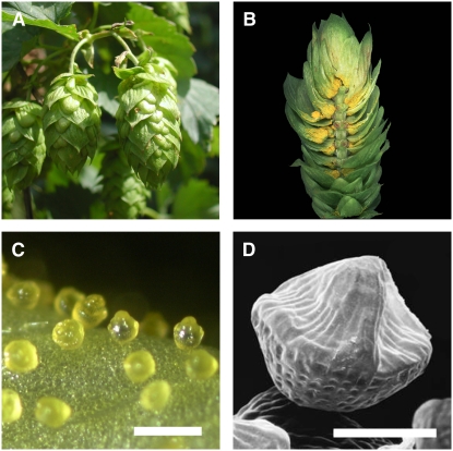
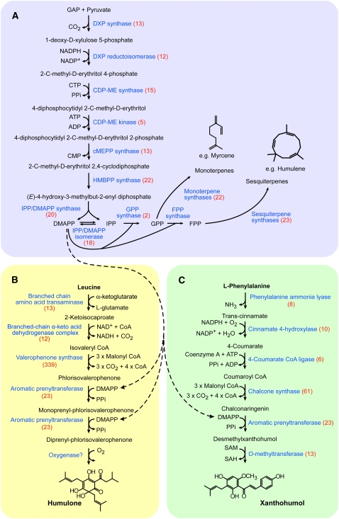
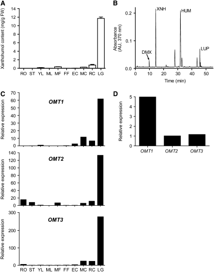
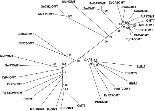
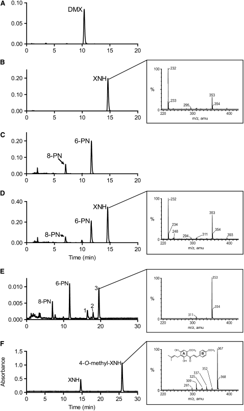
The glandular trichomes (lupulin glands) of hop (Humulus lupulus) synthesize essential oils and terpenophenolic resins, including the bioactive prenylflavonoid xanthohumol. To dissect the biosynthetic processes occurring in lupulin glands, we sequenced 10,581 ESTs from four trichome-derived cDNA libraries. ESTs representing enzymes of terpenoid biosynthesis, including all of the steps of the methyl 4-erythritol phosphate pathway, were abundant in the EST data set, as were ESTs for the known type III polyketide synthases of bitter acid and xanthohumol biosynthesis. The xanthohumol biosynthetic pathway involves a key O-methylation step. Four S-adenosyl-l-methionine-dependent O-methyltransferases (OMTs) with similarity to known flavonoid-methylating enzymes were present in the EST data set. OMT1, which was the most highly expressed OMT based on EST abundance and RT-PCR analysis, performs the final reaction in xanthohumol biosynthesis by methylating desmethylxanthohumol to form xanthohumol. OMT2 accepted a broad range of substrates, including desmethylxanthohumol, but did not form xanthohumol. Mass spectrometry and proton nuclear magnetic resonance analysis showed it methylated xanthohumol to 4-O-methylxanthohumol, which is not known from hop. OMT3 was inactive with all substrates tested. The lupulin gland-specific EST data set expands the genomic resources for H. lupulus and provides further insight into the metabolic specialization of glandular trichomes.


Figures

Figure 1.
Figure 1.
Morphology of Hop Cones and Lupulin Glands. (A) Cones of hop cultivar Taurus. Cones are ∼5 cm in length. (B) Longitudinal section of a hop cone showing lupulin glands at the base of bracteoles. (C) A light microscopy image of ripe lupulin glands. Bar = 500 μm. (D) Scanning electron micrograph of a ripe lupulin gland showing the peaked appearance of the filled subcuticular sac. Bar = 100 μm.
Figure 2.
Figure 2.
The Three Major Biosynthetic Pathways Active in Lupulin Glands. Enzyme names are shown in blue. Each enzyme is annotated with the number of corresponding ESTs shown in parentheses. Dashed arrows indicate the role of DMAPP for prenylation in the bitter acid and xanthohumol pathways. (A) The terpenoid pathway in hop lupulin glands. This pathway includes the MEP pathway for synthesis of IPP and DMAPP and also provides myrcene and humulene, the main terpenoids in hop lupulin glands. The mevalonate pathway is not shown because it appears to contribute little to the formation of lupulin gland terpenoids. (B) The bitter acid pathway in hop lupulin glands. The biosynthesis of humulone, the main α-acid found in hop trichomes, is shown. The nature of the aromatic prenylation and final oxidation steps are not known. (C) The xanthohumol pathway in hop lupulin glands. The aromatic prenyltransferase is not identified.
Figure 3.
Figure 3.
Quantification of Xanthohumol and Hl OMT Gene Expression Analysis of Different Hop Tissues. (A) Amounts of xanthohumol occurring in different tissues of the cultivar Taurus measured using HPLC. Values represent mean ± sd (n = 3). RO, roots; ST, stem; YL, young leaf; ML, mature leaf; MF, male flower; FF, female flower; EC, early-stage cone; MC, mid-stage cone; RC, ripe cone; LG, lupulin gland; FW, fresh weight. (B) A representative chromatogram obtained by HPLC analysis of a Taurus lupulin gland sample. DMX, desmethylxanthohumol; XNH, xanthohumol; HUM, humulone; LUP, lupulone. (C) Real-time PCR analysis of transcript levels of OMT1, OMT2, and OMT3 in Taurus tissues. Each sample contained pooled material from several collections of each tissue from multiple plants. Expression values were normalized with GAPDH amplification and are displayed relative to the expression level in young leaves. (D) Comparison of the relative expression of OMT1, OMT2, and OMT3 in lupulin glands using RT-PCR.
Figure 4.
Figure 4.
Unrooted Similarity Tree of Plant OMT Proteins Constructed Using the Neighbor-Joining Method. Bootstrap values from a minimum of 1000 trials are shown. Protein names and accession numbers are listed in Methods.
Figure 5.
Figure 5.
HPLC and LC-MS Analysis of Product Formation by Recombinant Hl OMT1 and OMT2. (A) Chromatogram of authentic desmethylxanthohumol (DMX) standard analyzed at an absorbance of 330 nm. (B) Chromatogram of authentic xanthohumol (XNH) standard at 330 nm. Inset: negative-ion ESI-LC-MS analysis of the xanthohumol standard. (C) HPLC analysis of the methylation of desmethylxanthohumol by OMT1 in the absence of the methyl donor SAM. The chromatogram was obtained at 330 nm. (D) HPLC analysis of the methylation of desmethylxanthohumol by OMT1 in the presence of SAM. OMT1 methylates desmethylxanthohumol to form xanthohumol (14.6 min). 6-PN and 8-PN are formed by isomerization of the substrate. The chromatogram was obtained at 330 nm. Inset: negative-ion ESI-LC-MS analysis of the 14.6 min peak produced by methylation of desmethylxanthohumol by OMT1. (E) HPLC analysis of the methylation of desmethylxanthohumol by OMT2. Desmethylxanthohumol spontaneously isomerizes to form 6-PN (11.7 min) and 8-PN (7.0 min) in the OMT assay. The identities of peaks 1 to 3 are described in Results. The chromatogram was extracted at 330 nm. Inset: negative-ion ESI-LC-MS analysis of the 19.5 min peak corresponding to a monomethylated prenylnaringenin-type flavanone (m/z 353). (F) HPLC analysis of the methylation of xanthohumol by OMT2. Inset: negative-ion ESI-LC-MS analysis of the product peak corresponding to 4-O-methylxanthohumol (m/z 367).

References

    1. Audic, S., and Claverie, J.M. (1997). The significance of digital gene expression profiles. Genome Res. 7 986–995. - PubMed
    1. Biendl, M. (2003 2002). Research on the xanthohumol content of hops. Hopfenrundschau International 2002/2003 72–75.
    1. Bonaldo, M.F., Lennon, G., and Soares, M.B. (1996). Normalization and subtraction: Two approaches to facilitate gene discovery. Genome Res. 6 791–806. - PubMed
    1. Burga, L., Wellmann, F., Lukacin, R., Witte, S., Schwab, W., Schroder, J., and Matern, U. (2005). Unusual pseudosubstrate specificity of a novel 3,5-dimethoxyphenol O-methyltransferase cloned from Ruta graveolens L. Arch. Biochem. Biophys. 440 54–64. - PubMed
    1. Burke, C.C., Wildung, M.R., and Croteau, R. (1999). Geranyl diphosphate synthase: Cloning, expression, and characterization of this prenyltransferase as a heterodimer. Proc. Natl. Acad. Sci. USA 96 13062–13067. - PMC - PubMed

Publication types

MeSH terms

Associated data