

PHACES association: a neuroradiologic review of 17 patients
- PMID: 18223093
- PMCID: PMC7978195
- DOI: 10.3174/ajnr.A0937
PHACES association: a neuroradiologic review of 17 patients
Abstract
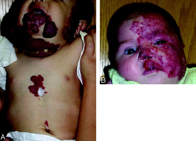
Background and purpose: We present neuroradiologic findings in 17 patients with posterior fossa malformations, hemangiomas, arterial anomalies, cardiac defects, eye abnormalities, and sternal or ventral defects (PHACES) association and identify those at highest risk of central nervous system (CNS) structural, cerebrovascular, and neurodevelopmental abnormalities.
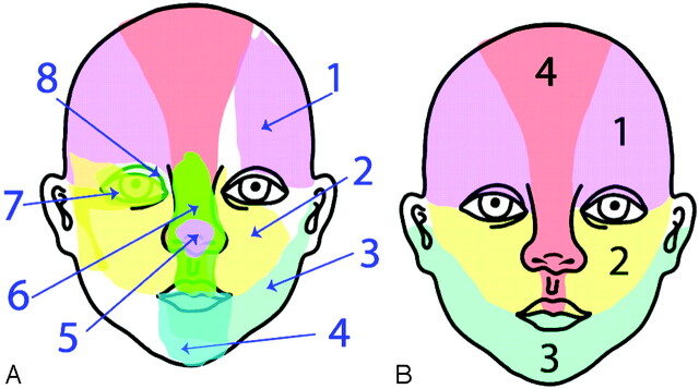
Materials and methods: Patients with PHACES association were identified in the Vascular Anomalies Program at New York University Medical Center from 1998 to 2007. Many patients were followed in conjunction with other specialists at the Birthmark Institute at Roosevelt Hospital. Clinical records and imaging studies were reviewed retrospectively. Criteria for diagnosis of PHACES were based on previously published indicators. Imaging studies were independently re-reviewed by a neuroradiologist. Segmental mapping of cutaneous hemangioma distribution by photograph review and presence or absence of other PHACES-associated findings were correlated with radiologic findings.
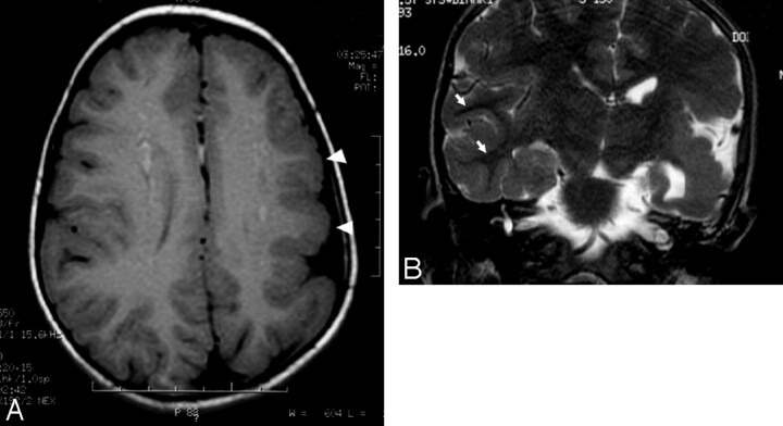
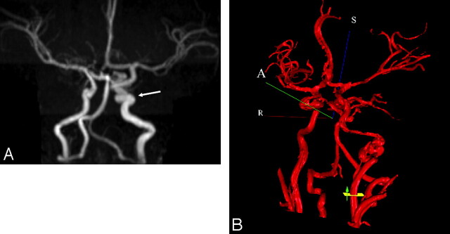
Results: Patients with large facial cutaneous (S1-S4) hemangiomas were especially at risk of CNS structural and cerebrovascular anomalies; S1 with ocular anomalies; and S3 with airway, ventral, and cardiac anomalies. All patients with CNS structural malformations had a cerebrovascular abnormality, and this cohort was at risk for developmental and/or other neurologic sequelae. Four patients had supratentorial CNS anomalies, including cortical dysgenesis and migration abnormalities. Some patients with CNS arteriopathy progressed to aneurysms.
Conclusion: Our data support and expand the work of others, identifying risk factors for segmental hemangiomas. In addition to posterior fossa CNS anomalies, supratentorial anomalies may be present in patients with PHACES, and this may correlate with significant clinical sequelae. The long-term prognosis of these patients remains unknown.
Figures
Comment in
-
PHACES syndrome: from the brain to the face via the neural crest cells.AJNR Am J Neuroradiol. 2008 Apr;29(4):814-5. doi: 10.3174/ajnr.A0943. Epub 2008 Jan 17. AJNR Am J Neuroradiol. 2008. PMID: 18202231 Free PMC article. No abstract available.
References
-
- Haggstrom AN, Lammer EJ, Schneider RA, et al. Patterns of infantile hemangiomas: new clues to hemangioma pathogenesis and embryonic facial development. Pediatrics 2006;117:698–703 - PubMed
-
- Waner M, North PE, Scherer KA, et al. The nonrandom distribution of facial hemangiomas. Arch Dermatol 2003;139:869–75 - PubMed
-
- Pascual-Castroviejo I. Vascular and nonvascular intracranial malformation associated with external capillary hemangiomas. Neuroradiology 1978;16:82–84 - PubMed
-
- Frieden IJ, Reese V, Cohen D. PHACE syndrome: the association of posterior fossa brain malformations, hemangiomas, arterial anomalies, coarctation of the aorta and cardiac defects, and eye abnormalities. Arch Dermatol 1996;132:307–11 - PubMed
-
- Pascual-Castroviejo I, Pascual-Pascual SI, Rafia S, et al. Hemangiomas, and cutaneous and intracranial vascular deformations (Pascual-Castroviejo syndrome type II): a case report [in Spanish]. Rev Neurol 2002;35:1034–36 - PubMed
MeSH terms
LinkOut - more resources
Full Text Sources