. 2008 Mar;7(3):509-17.
doi: 10.1128/EC.00316-07. Epub 2008 Jan 25.

Glycogen phosphorylase in Acanthamoeba spp.: determining the role of the enzyme during the encystment process using RNA interference

Affiliations

Glycogen phosphorylase in Acanthamoeba spp.: determining the role of the enzyme during the encystment process using RNA interference

Jacob Lorenzo-Morales et al. Eukaryot Cell. 2008 Mar.

Abstract

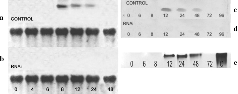
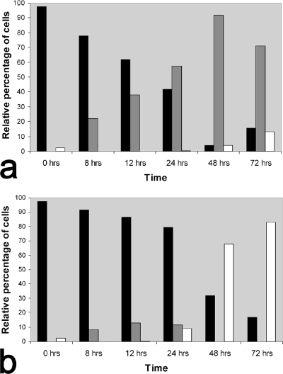
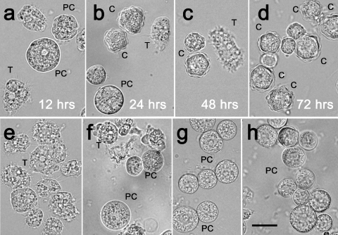
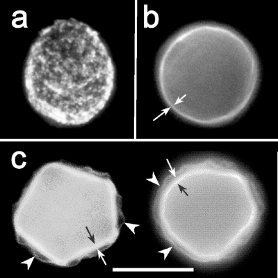
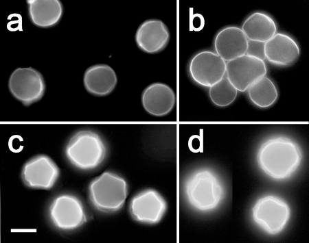
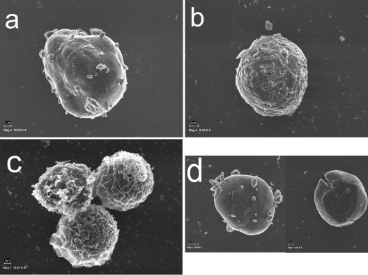
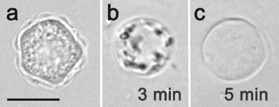
Acanthamoeba infections are difficult to treat due to often late diagnosis and the lack of effective and specific therapeutic agents. The most important reason for unsuccessful therapy seems to be the existence of a double-wall cyst stage that is highly resistant to the available treatments, causing reinfections. The major components of the Acanthamoeba cyst wall are acid-resistant proteins and cellulose. The latter has been reported to be the major component of the inner cyst wall. It has been demonstrated previously that glycogen is the main source of free glucose for the synthesis of cellulose in Acanthamoeba, partly as glycogen levels fall during the encystment process. In other lower eukaryotes (e.g., Dictyostelium discoideum), glycogen phosphorylase has been reported to be the main tool used for glycogen breakdown in order to maintain the free glucose levels during the encystment process. Therefore, it was hypothesized that the regulation of the key processes involved in the Acanthamoeba encystment may be similar to the previously reported regulation mechanisms in other lower eukaryotes. The catalytic domain of the glycogen phosphorylase was silenced using RNA interference methods, and the effect of this phenomenon was assessed by light and electron microscopy analyses, calcofluor staining, expression zymogram assays, and Northern and Western blot analyses of both small interfering RNA-treated and control cells. The present report establishes the role of glycogen phosphorylase during the encystment process of Acanthamoeba. Moreover, the obtained results demonstrate that the enzyme is required for cyst wall assembly, mainly for the formation of the cell wall inner layer.


Figures

FIG. 1.
FIG. 1.
Results from agarose gel electrophoresis showing PCR amplifications with the AGP primer pair. Lanes: 1, 100-bp DNA ladder; 2, A. astronyxis; 3, SWT-22; 4, MN-7; 5, A. castellanii Neff; 6, empty; 7, negative control.
FIG. 2.
FIG. 2.
Analyses of glycogen phosphorylase expression during encystation. (a and b) Northern blot analysis of glycogen phosphorylase expression during encystation in siRNA-treated (b) and control (a) cultures of Acanthamoeba. Total RNA extracted at different time points during encystation was electrophoresed at 80 V, blotted onto a nylon membrane, and probed with cDNA for glycogen phosphorylase. Top lanes, total RNA hybridized with cDNA corresponding to the catalytic domain of Acanthamoeba glycogen phosphorylase. Bottom lanes, hybridization with 18S mRNA as a control. Numbers indicate the hours after the induction of encystation. (c and d) Western blot analysis of glycogen phosphorylase during encystation in siRNA-treated (d) and control (c) cultures of Acanthamoeba. Protein extracts from different time points during encystation were incubated with polyclonal anti-human glycogen phosphorylase (BB) rabbit antibody, and results were developed with digoxigenin. Numbers indicate the hours after the induction of encystation. (e) Analysis of glycogen phosphorylase activity during encystation of Acanthamoeba. Proteins were isolated in time course experiments and separated by nondenaturing PAGE on a gel containing glycogen. Zymograms were developed by iodine staining of polyglucan synthesized from glucose-1-phosphate. Numbers indicate the hours after the induction of encystation. Lane C, positive control with glycogen phosphorylase b from rabbit muscle.
FIG. 3.
FIG. 3.
Distribution of trophozoites, immature precysts, and mature cysts during the course of encystment in the presence (a) or absence (b) of glycogen phosphorylase siRNA. The cell distribution (percentages of different cell types at different time points after the induction of encystation) was determined by counting cells viewed under an inverse microscope. The minimum number of cells counted per time point was 130; the maximum number of cells counted per time point was 600. All experiments were repeated five times. Black columns, trophozoites; gray columns, immature precysts; white columns, mature cysts.
FIG. 4.
FIG. 4.
Morphology of the living acanthamoebae at different time points after the induction of encystation. (a to d) Control culture. (a) Twelve hours after the induction of encystation, the trophozoites are detaching from the flask and starting to round up. (b) Twenty-four hours after the induction of encystation, immature cysts with a single-layered cyst wall are present; the first mature cysts with both layers of the cyst wall are also shown. (c and d) From 48 to 72 h after the induction of encystation, the number of mature cysts with both walls gradually increases, and at 72 h, the population of acanthamoebae consists mainly of mature cysts. (e to h) siRNA-treated cultures. (e) Twelve hours after the induction of encystation, trophozoites are starting to round up, similar to those in the control culture. (f) Twenty-four hours after the induction of encystation, single-layered “pseudocysts” are observed as a main cell type. (g) No mature cysts are observed 48 h after the induction of encystation. The main cell type present is the single-layered immature cyst. (h) Immature cysts represent approximately 70% of the cell population; the first mature cysts are also observed. T, trophozoite; PC, immature precyst; C, mature cyst. Scale bar, 10 μm.
FIG. 5.
FIG. 5.
Phases of cyst wall formation as detected by calcofluor staining. (a) The rounded trophozoite with cellulose patches on the cell surface appears approximately 12 h after the induction of encystation. (b) Immature precyst with a continuous cell wall containing cellulose (approximately 24 h after the induction of encystation). A single layer of the cell wall is indicated by arrows. (c) Two mature cysts with both layers of the cell wall: a wrinkled exocyst (arrows) and an endocyst (arrowheads) containing a larger amount of cellulose than the exocyst.
FIG. 6.
FIG. 6.
Cyst types characteristic of stages of Acanthamoeba encystment in the presence or absence of siRNA for glycogen phosphorylase. (a and b) siRNA-treated culture after 24 h (a) or 48 h (b) of encystment. (c and d) Untreated control culture after 24 h (c) or 48 h (d) of encystment. The single-layered immature precysts represented the main cell type in the siRNA-treated cultures at both time points, whereas in the untreated cultures, the mature cysts with both cyst wall layers were already present after 24 h and prevailed after 48 h of encystment. Scale bar, 10 μm.
FIG. 7.
FIG. 7.
Phases of cyst wall formation as detected by scanning electron microscopy. (a) The rounded trophozoite with cellulose patches on the cell surface appears approximately 12 h after the induction of encystation. (b) Immature precyst with a continuous cell wall (approximately 24 h after the induction of encystation). A single layer of the cell wall is indicated by arrows. (c) Three mature cysts. The wrinkled exocyst is clearly recognizable. (d) RNAi-treated acanthamoebae were not able to form a mature cyst. Mag, magnification; 12.00 K X, ×12,000.
FIG. 8.
FIG. 8.
Sensitivity to 0.5% SDS of 72-h-old cyst stages formed in siRNA-treated and untreated cultures. (a) The mature cyst from the control cultures shows no affected morphology after a 5-min exposure to SDS. Scale bar, 10 μm. (b) The immature precyst from the siRNA-treated culture is undergoing lysis after a 3-min treatment with SDS. (c) The empty single-layered cyst wall shelter is a residuum of the SDS-treated precyst.

References

    1. Anderson, I. J., R. F. Watkins, J. Samuelson, D. F. Spencer, W. H. Majoros, M. W. Gray, and B. J. Loftus. 2005. Gene discovery in the Acanthamoeba castellanii genome. Protist 156203-214. - PubMed
    1. Barrett, R. A., and M. Alexander. 1977. Resistance of cysts of amoebae to microbial decomposition. Appl. Environ. Microbiol. 33670-674. - PMC - PubMed
    1. Bishop, J. D., B. C. Moon, F. Harrow, D. Ratner, R. H. Gomer, R. P. Dottin, and D. T. Brazill. 2002. A second UDP-glucose pyrophosphorylase is required for differentiation and development in Dictyostelium discoideum. J. Biol. Chem. 3632430-32437. - PubMed
    1. Blanton, W. E., and C. L. Villemez. 1978. Molecular-size and chain-length distribution in Acanthamoeba cellulose. J. Protozool. 25264-267.
    1. Booton, G. C., D. J. Kelly, Y. W. Chu, D. V. Seal, E. Houang, D. S. Lam, T. J. Byers, and P. A. Fuerst. 2002. 18S ribosomal DNA typing and tracking of Acanthamoeba species isolates from corneal scrape specimens, contact lenses, lens cases, and home water supplies of Acanthamoeba keratitis patients in Hong Kong. J. Clin. Microbiol. 401621-1625. - PMC - PubMed

Publication types

Associated data

LinkOut - more resources