. 2008 Mar;237(3):592-601.
doi: 10.1002/dvdy.21439.

The emergence of ectomesenchyme

Affiliations

The emergence of ectomesenchyme

Aida Blentic et al. Dev Dyn. 2008 Mar.

Abstract

In the head, neural crest cells generate ectomesenchymal derivatives: cartilage, bone, and connective tissue. Indeed, these cells generate much of the cranial skeleton. There have, however, been few studies of how this lineage is established. Here, we show that neural crest cells stop expressing early neural crest markers upon entering the pharyngeal arches and switch to become ectomesenchymal. By contrast, those neural crest cells that do not enter the arches persist in their expression of early neural crest markers. We further show that fibroblast growth factor (FGF) signaling is involved in directing neural crest cells to become ectomesenchymal. If neural crest cells are rendered insensitive to FGFs, they persist in their expression of early neural crest markers, even after entering the pharyngeal arches. However, our results further suggest that, although FGF signaling is required for the realization of the ectomesenchymal lineages, other cues from the pharyngeal epithelia are also likely to be involved.


Figures

Figure 1
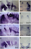
Figure 1. Disposition of ectomesenchymal and non-ectomesenchymal crest within the cranial neural crest
Ap2α is a general marker of the cranial neural crest, labelling neural crest within the pharyngeal arches as well as those lying between the arches and the hindbrain (a, b). Sox10 (c,d) and Foxd3 (e,f) are expressed by the crest that lie between the arches and the neural tube. Sox10 is additionally expressed in otic vesicle (c,d). The ectomsenchymal crest can be identified through their expression of Dlx2 (g,h). Dlx2 only labels the crest cells within the pharyngeal arches. (a,c,e,g) Lateral view of the head of stage 15 chick embryos with anterior to the left and dorsal to the top. (b,d,f,h) Transverse sections (40μm), shown at the level of otic vesicle of stage 19 embryos., with dorsal to the top ov, otic vesicle; I,II,III,IV the position of each pharyngeal arches.
Figure 2
Figure 2. Emigrant neural crest cells are non-ectomesenchymal in nature
Cranial neural crest cells emerging from the midbrain and anterior hindbrain at HH10 express Sox-10 (a) and Foxd3 (d). At this stage Dlx2 is not expressed by any neural crest cells, although this gene is expressed in rhombomere 5 of the hindbrain. At stage 11, neural crest cells emerge from rhombomeres 4 and 6 of the hindbrain, express Sox-10 (b) and Foxd3 (e). Dlx2 is still not expressed by neural crest cells, although there continues to be some expression in rhombomere 5 and also now in the pharyngeal endoderm. By stage 14 of development, Sox10 (c) and Foxd3 (f) expression is restricted to the dorsally located crest, while the more ventral crest that has started to populate the pharyngeal arches now expresses Dlx2 (i) (a,b,d,e,g,h) Dorsal views of the neural tube with anterior to the left and posterior to the right. (c,f,i) Lateral view of pharyngeal arches with anterior to the left, dorsal to the top. op, otic placode; r2, rhombomere 2, r4, rhombomere 4, r5, rhombomere 5; other abbreviations as before.
Figure 3
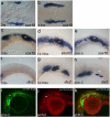
Figure 3. Appearance of non-ectomesenchymal and ectomesenchymal crest in the zebrafish
a: Neural crest cells can be first identified alongside the neural tube at the midbrain level at the 3 somite stage through their expression of sox10. b,c: As development proceeds, sox10 expression is associated with neural crest cells from more posterior regions. f: At the 12 somite stage, dlx2 is expressed in the forebrain but not in neural crest cells. d,g: At 14 somites, sox10 expression is restricted to those crest cells that lie dorsally, close to the neural tube (d), while dlx2 can now be seen in the ectomesenchymal crest that have filled the first three pharyngeal arches (g). e,h: By the prim-5 stage, sox10 expression is still restricted to the more dorsal crest (e) and dlx2 to the ectomesenchymal crest of the first four pharyngeal arches (h). c-e: From the 12 somite stage onwards, sox10 expression is associated with the developing inner ear. To test the hypothesis that ectomesenchymal crest emerge from within the ectomesenchymal crest, we have analysed the distribution of GFP protein and GFP RNA in a sox10 transgenic reporter line. i-k: GFP protein is found in all the cranial neural crest cells (i, k) but GFP RNA is only found in the non-ectomesenchymal crest (j). (k) Overlay GFP protein with GFP RNA. In all panels, anterior is to the left and dorsal to the top. Abbreviations as before.
Figure 4
Figure 4. Dlx2 expression is absent from zebrafish embryos treated with an inhibitor of FGF signalling
In embryos treated with DMSO alone, sox10 expression is found in the dorsally located crest (a) and dlx2 in the ectomesenchymal crest within the pharyngeal arches (c). In embryos treated with SU5402, sox10 expressing crest are still found in the more dorsal position (b), but there is a lack of dlx2 expression (d). There seems to be an abundance of post-otic sox10 expressing crest in the SU5402 treated embryos (c). (a-d) Lateral view of prim-5 embryos. In all panels, anterior is to the left and dorsal to the top. Abbreviations as before.
Figure 5
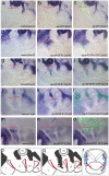
Figure 5. Neural crest cells insensitive to FGF signalling persist in their expression of non-ectomesenchymal markers even after they have entered the pharyngeal arches
(a-c) Embryos electroporated with pCAβ-eGFP at 9 somite stage (HH9+). Normal expression of Sox10 is seen on both the non-electroporated side (a) and also on the electroporated side (b). (c) Overlay with GFP positive cells. (d-f) Embryo electroporated with dnFGFR1-GFP at the 8 somite stage (HH9-). (d) non-electoporated/control side shows normal Sox10 expression. Contrstaingly, ectopic Sox10 expression is seen in cells that have entered the first arch on the electroporated side (e). (f) Overlay of the Sox10 expression with the dnFGFR1-GFP positive cells in green. (g-i) Embryos electroporated with dnFGFR1-GFP at the 9 somite stage (HH9+). (g) non-electroporated control side shows normal Sox10 expression. (h) On the electroporated side, ectopic Sox10 expression can be seen in the second arch. (i) Overlay of Sox10 expression with dnFGFR1-GFP positive cells in green. (j-l) Embryos electroporated with dnFGFR1-GFP at the 6 somite stage (HH9-). (j) non-electroporated control side shows normal Foxd3 expression. (k) On the electroporated side, ectopic Foxd3 expression can be seen in the second arch. (l) Overlay of Foxd3 expression with dnFGFR1-GFP positive cells in green. (m-o) Embryos electroporated with dnFGFR1-GFP at the 9 somite stage (HH9+). (m) non-electroporated control side shows normal Dlx2 expression. (n) On the electroporated side, diminished Dlx2 expression can be seen in the second arch. (i) Overlay of Dlx2 expression with dnFGFR1-GFP positive cells in green. Schematic illustration of cranial neural crest at the level of pharyngeal arches at HH15, normal distribution of non-ectomesenchymal crest (p). Ectopic non-ectomesenchymal crest cells are seen within the pharyngeal arches when crest cells carry the domninant negative FGF receptorin (q, r). This is shown in side view (q) and in transverse (r). In the tranverse view (r) ectopic ectomesenchymal crest cells are only seen in the electroprated right side. In panels (a - q), anterior is to the left and dorsal to the top. In (r), dorsal is to the top.
Figure 6
Figure 6. Expression of members of the FGF synexpression group during the period of neural crest migration
Dorsal views of stage 11 embryos, anterior to the left, showing (a) erm expression (b) pea3 expression (c) sprouty 2 expression and (d) dpERK immostaining. In all cases, expression of erm, pea3, sprouty2 and dpERK staining can be seen at the anterior neuropore, around the isthmus in the mesencephalon and anterior hindbrain, and, with the exception of sprouty 2, in the central region of the hindbrain and in the otic placode. Expression is not seen in the neural crest cells, which at this stage are in abundance lateral to the neural tube (see Fig 2). AN - anterior neuropore, Mes - mesencephalon, I - Isthmus, Hind - Hindbrain, Op - Otic placode.

References

    1. Abzhanov A, Tzahor E, Lassar AB, Tabin CJ. Dissimilar regulation of cell differentiation in mesencephalic (cranial) and sacral (trunk) neural crest cells in vitro. Development. 2003;130:4567–4579. - PubMed
    1. Baker CV, Bronner-Fraser M, Le Douarin NM, Teillet MA. Early- and late-migrating cranial neural crest cell populations have equivalent developmental potential in vivo. Development. 1997;124:3077–3087. - PubMed
    1. Britsch S, Goerich DE, Riethmacher D, Peirano RI, Rossner M, Nave K-A, Birchmeier C, Wegner M. The transcription factor Sox10 is a key regulator of peripheral glial development. Genes Dev. 2001;15:66–78. - PMC - PubMed
    1. Carney TJ, Dutton KA, Greenhill E, Delfino-Machin M, Dufourcq P, Blader P, Kelsh RN. A direct role for Sox10 in specification of neural crest-derived sensory neurons. Development. 2006 - PubMed
    1. Chambers D, Mason I. Expression of sprouty2 during early development of the chick embryo is coincident with known sites of FGF signalling. Mech Dev. 2000;91:361–364. - PubMed

Publication types

LinkOut - more resources