. 2008 Mar;237(3):630-9.
doi: 10.1002/dvdy.21446.

Cellular and molecular determinants targeting the Caenorhabditis elegans PHR protein RPM-1 to perisynaptic regions

Affiliations

Cellular and molecular determinants targeting the Caenorhabditis elegans PHR protein RPM-1 to perisynaptic regions

Benjamin Abrams et al. Dev Dyn. 2008 Mar.

Abstract

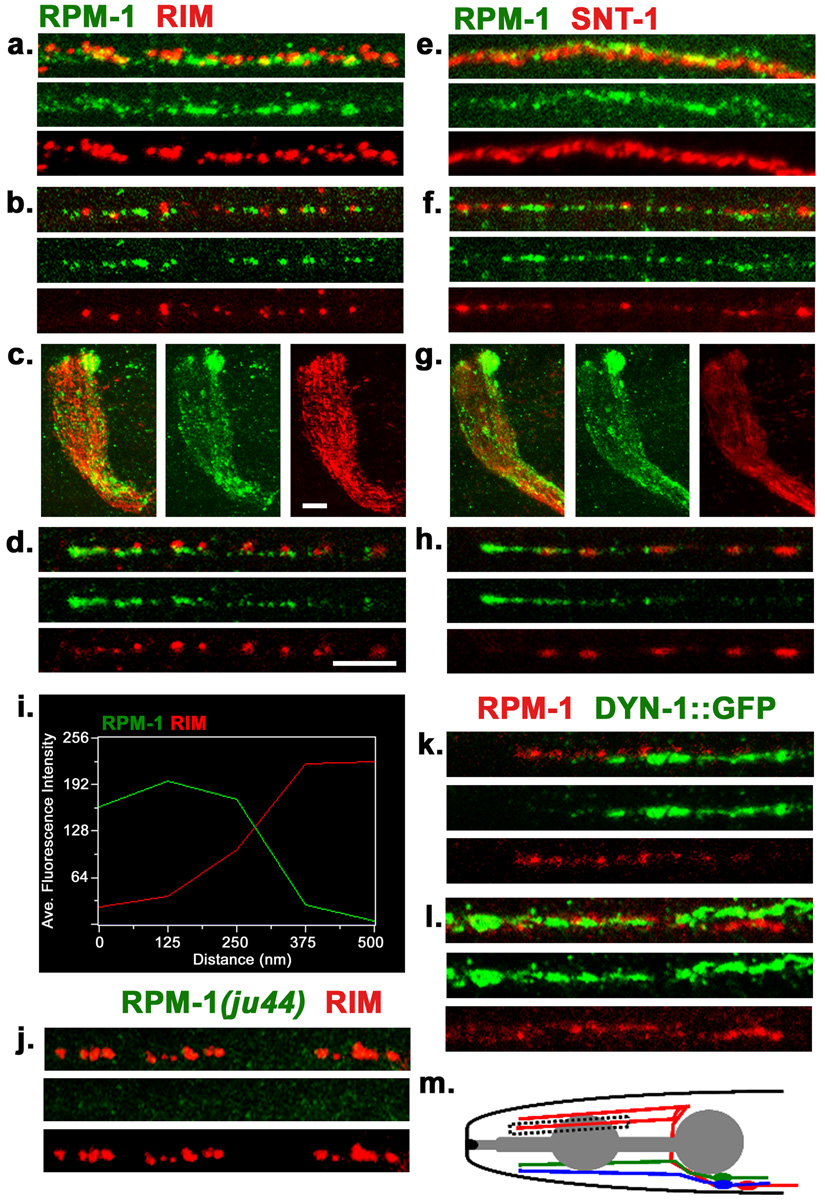
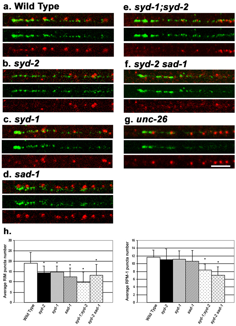
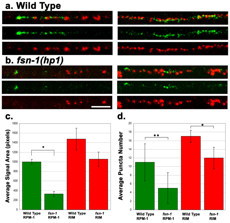
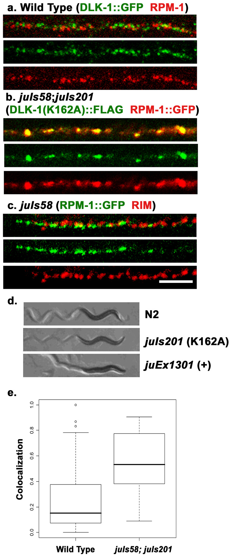
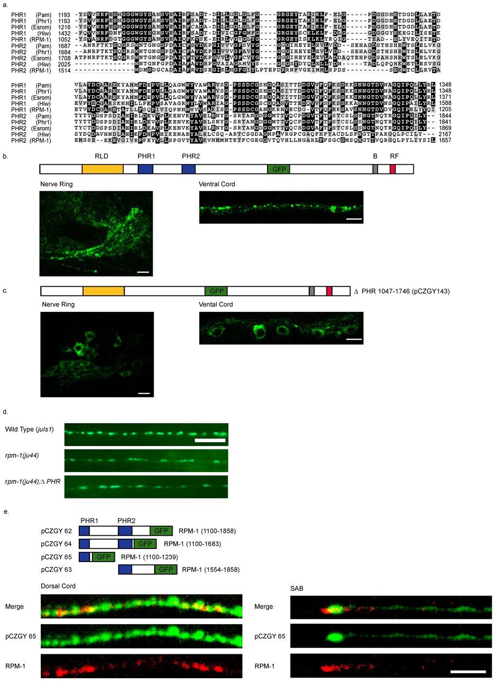
Caenorhabditis elegans RPM-1 is a member of a conserved protein family, the PHR proteins, that includes human Pam, mouse Phr1, zebrafish Esrom, and Drosophila Highwire. PHR proteins play important roles in the development of the nervous system. In particular, mutations in rpm-1 cause a disruption of synaptic architecture, affecting the distribution of synaptic vesicles and the number of presynaptic densities. Using antibodies against RPM-1, we determined the localization of the endogenous RPM-1 protein in wild-type and in several mutants that affect synaptic development. Our analyses show that, in mature neurons, RPM-1 resides in a distinct region that is close to, but does not overlap with, the synaptic exo- and endocytosis domains. The localization of RPM-1 occurs independently of several proteins that function in the transport or assembly of synapse components, and its abundance is partially dependent on its binding partner the F-box protein FSN-1. RPM-1 has been shown to target the MAPKKK DLK-1 for degradation. We show that activated DLK-1 may be preferentially targeted for degradation. Furthermore, using transgene analysis, we identified a critical role of the conserved PHR domain of RPM-1 in its subcellular localization.


Figures

Figure 1
Figure 1. Endogenous localization of RPM-1
(a–d) Confocal images of wild type (N2) worms co-stained with anti-RPM-1 (green) and anti-RIM (red) in (a) dorsal cord, (b) sub-lateral cord, (c) nerve ring, (d) SAB neuron. (e–h) Confocal images of N2 worms co-stained with RPM-1 (green) and synaptotagmin/SNT-1 (red) in (e) dorsal cord, (f) sub-lateral cord, (g) nerve ring, (h) SAB neuron. (i) Fluorescence intensity plot showing the typical distance between RPM-1 puncta and RIM in the dorsal cord. (j) The RPM-1 antibody is specific. Confocal images of the dorsal cord of rpm-1(ju44) mutants. RIM stain (red) is positive and shows gaps that are characteristics of the Rpm-1 mutant phenotype, whereas no RPM-1 staining is detected. (k–l) juIs83 (DYNAMIN-1::GFP) worms co-stained with RPM-1(red) and GFP (green) in (k) SAB neuron and (l) dorsal cord. (m) Schematic drawing of the SAB neurons. Blue, SABVL; green, SABVR; red, SABD. All SABs have their cell bodies in the retro-vesicular ganglion, project axons towards the anterior, and form en passant synapses onto head muscles. Box indicates region of SAB shown in d and h. All SAB images shown are in the same orientation with the anterior end on the left, individual SAB processes were chosen at random. Scale bars: 5µm, a, b, d, e, f, h, k, l, j . Scale bar, 10µm c, g,
Figure 2
Figure 2. RPM-1 localization in mutants that have altered synaptic morphology
Shown are confocal images of SAB neurons co-stained with RPM-1 (green) and RIM (red). SAB regions are same as indicated in figure 1m. (a) Wild type (N2). (b) syd-2(ju37) (c) syd-1(ju82) (d) sad-1(ju53) (e) syd-1(ju82);syd-2(ju37) (f) syd-2(ju37)sad-1(ju53) (g) unc-26(e1196). In syd-1 and syd-2 mutants, RIM expression is diffuse and reduced in intensity, but RPM-1 expression is normal. (h) Quantitation of RIM (left plot) or RPM-1 (right plot) puncta number per 20µm length of dorsal cord. t-tests were performed for RPM-1 and RIM puncta number relative to wild type, * p<0.05. Scale bar, 5µm, all panels are the same scale.
Figure 3
Figure 3. RPM-1 expression is reduced in fsn-1(hp1) mutants
(a–b) Shown are confocal images of SAB neurons (left panels) or dorsal cord (right panels) co-stained with RPM-1 (green) and RIM (red). (a) N2 (wild type), a gradient of RPM-1 staining is observed in the SAB neurons that is brightest near the anterior tip of SAB axon, and tapers off in the posterior part of the axon. Dorsal cord regions do not show a gradient pattern. (b) In fsn-1(hp1) mutants, the abundance of RPM-1 is significantly decreased in the SAB and dorsal cord, though the RPM-1 staining is still brightest at the anterior end of the SAB. (c) Quantitation of RPM-1 staining abundance. Bar graph shows the average total number of pixels that contained above background signal for a section of dorsal cord, with error bars as std. dev. RPM-1 abundance is significantly decreased in fsn-1(hp1) animals, green bars *(p<10−6). RIM abundance is not significantly different from wild type, red bars (p=0.09). n = 6 dorsal cords for each data set. P-value is for a Welch Two Sample t-test. (d) Quantitation of puncta number shows a greater decrease in RPM-1 compared to RIM (for RPM-1, P<10−4; for RIM, p=0.02). Scale bar 5µm, same scale for all panels.
Figure 4
Figure 4. Colocalization studies of RPM-1 and DLK-1
(a) Confocal image of the dorsal cord of juIs192[DLK-1::GFP] animals co-stained with GFP (green) for DLK-1::GFP and endogenous RPM-1 (red). (a). In wild type animals expressing DLK-1::GFP at a low level that does not cause detectable synaptic defects, RPM-1 and DLK-1::GFP are seen in close proximity and rarely overlap. (b) Confocal image of juIs58 (RPM-1::GFP); juIs201[ DLK-1(K162A)::FLAG] animals, co-stained with anti-GFP (red) and anti-FLAG (green). the kinase dead DLK-1 shows extensive colocalization with RPM-1. (c) In juIs58 animals, RPM-1::GFP (green) shows normal spatial relationship to RIM (red). (d) Shown are bright-field images of animals and their movement trails. Wild type animals move sinusoidally. Over expression of wild type DLK-1 (juEx1301) causes animals to be hyper-contracted and uncoordinated (bottom). Overexpression of the kinase dead DLK-1 (K162A)(juIs201) does not have any effects on locomotion (middle). (e) Quantitation of the amount of DLK-1 signal present per RPM-1 puncta. Colocalization is significantly increased in juIs58;juIs201 animals compared to wild type, but is not complete. Colocalization is the ratio of DLK-1 signal area within RPM-1 signal area/puncta (see methods). N = 34 RPM-1 puncta per genotype, p<0.0001. Scale bar 5 µm, same scale for a–c.
Figure 5
Figure 5. Identification of domains important for RPM-1 localization
Constructs expressing RPM-1 or RPM-1::GFP are shown above the images. (a) Schematic of RPM-1 protein showing RCC1-like Domain (RLD), Pam, HIW ,RPM-1 (PHR1 and PHR2) domains, B-Box Zn finger domain (B) and RING-finger domain (RF). Dashed line in top panel of (a) indicates region removed in lower panel of (a). Confocal images in (a) show the dorsal cord in animals co-stained with RPM-1 (green) and RIM (red). The genotypes are wild type (top) and rpm-1(ju23);juEx743. Removal of residues 1862 to 3021 (dashed line) of RPM-1 abolishes RPM-1 activity but does not alter its spatial relationship to RIM. (b) Confocal images of GFP fluorescence in sub-lateral cords of transgenic animals expressing RPM-1::GFP. Number indicates the amino acid residue where RPM-1 is truncated and GFP is fused. Note that truncations all localize similarly to full length, until the first PHR domain is eliminated. Expression of RPM-1(1–851)::GFP is very diffuse and barely detectable in the sub-lateral cord (top), but is seen in the cell body in ventral cord (bottom). Scale bar 5 µm for all panels.
Figure 6
Figure 6. The PHR domain of RPM-1 is necessary and sufficient for synaptic localization
(a) A CLUSTAL alignment of the PHR regions. (b–c) Confocal images of GFP fluorescence in transgenic animals. (b) Expression of the functional full-length RPM-1::GFP(juIs58) is primarily in the nerve ring and nerve cords, and barely detectable in the cell bodies. (c) Deletion of the PHR domains juEx143 [pCZGY143] causes GFP to be confined to the cell bodies and is barely seen in nerve processes. (d) Expression of RPM-1ΔPHR::GFP (pCZGY143) does not rescue rpm-1(ju44). Wild type animals expressing juIs1[P unc-25 SNB-1::GFP] show an array of evenly sized, evenly spaced fluorescent puncta (top). rpm-1(ju44) animals display enlarged puncta with gaps in SNB-1GFP expression (middle), and are not rescued by the expression of RPM-1ΔPHR::GFP (pCZGY143) (bottom). (e) Schematics of the constructs expressing regions of PHR fused to GFP. The images are juEx1172 [pCZGY65] (pan neural PHR1::GFP) co-stained with endogenous RPM-1 and GFP in the dorsal cord (left panels) and in the SAB neurons (right panels). Scale bars 5µm.

References

    1. Ackley BD, Harrington RJ, Hudson ML, Williams L, Kenyon CJ, Chisholm AD, Jin Y. The Two Isoforms of the Caenorhabditis elegans Leukocyte-Common Antigen Related Receptor Tyrosine Phosphatase PTP-3 Function Independently in Axon Guidance and Synapse Formation. J. Neurosci. 2005;25:7517–7528. - PMC - PubMed
    1. Bloom AJ, Miller BR, Sanes JR, DiAntonio A. The requirement for Phr1 in CNS axon tract formation reveals the corticostriatal boundary as a choice point for cortical axons. Genes Dev. 2007;21:2593–2606. - PMC - PubMed
    1. Burgess RW, Peterson KA, Johnson MJ, Roix JJ, Welsh IC, O'Brien TP. Evidence for a conserved function in synapse formation reveals Phr1 as a candidate gene for respiratory failure in newborn mice. Mol Cell Biol. 2004;24:1096–1105. - PMC - PubMed
    1. Coyle IP, Koh YH, Lee WC, Slind J, Fergestad T, Littleton JT, Ganetzky B. Nervous wreck, an SH3 adaptor protein that interacts with Wsp, regulates synaptic growth in Drosophila. Neuron. 2004;41:521–534. - PubMed
    1. Crump JG, Zhen M, Jin Y, Bargmann CI. The SAD-1 kinase regulates presynaptic vesicle clustering and axon termination. Neuron. 2001;29:115–129. - PubMed

Publication types

MeSH terms

LinkOut - more resources