. 2008 Jan 28:8:26.
doi: 10.1186/1471-2148-8-26.

Evolution of a family of metazoan active-site-serine enzymes from penicillin-binding proteins: a novel facet of the bacterial legacy

Affiliations

Evolution of a family of metazoan active-site-serine enzymes from penicillin-binding proteins: a novel facet of the bacterial legacy

Nina Peitsaro et al. BMC Evol Biol. .

Abstract

Background: Bacterial penicillin-binding proteins and beta-lactamases (PBP-betaLs) constitute a large family of serine proteases that perform essential functions in the synthesis and maintenance of peptidoglycan. Intriguingly, genes encoding PBP-betaL homologs occur in many metazoan genomes including humans. The emerging role of LACTB, a mammalian mitochondrial PBP-betaL homolog, in metabolic signaling prompted us to investigate the evolutionary history of metazoan PBP-betaL proteins.

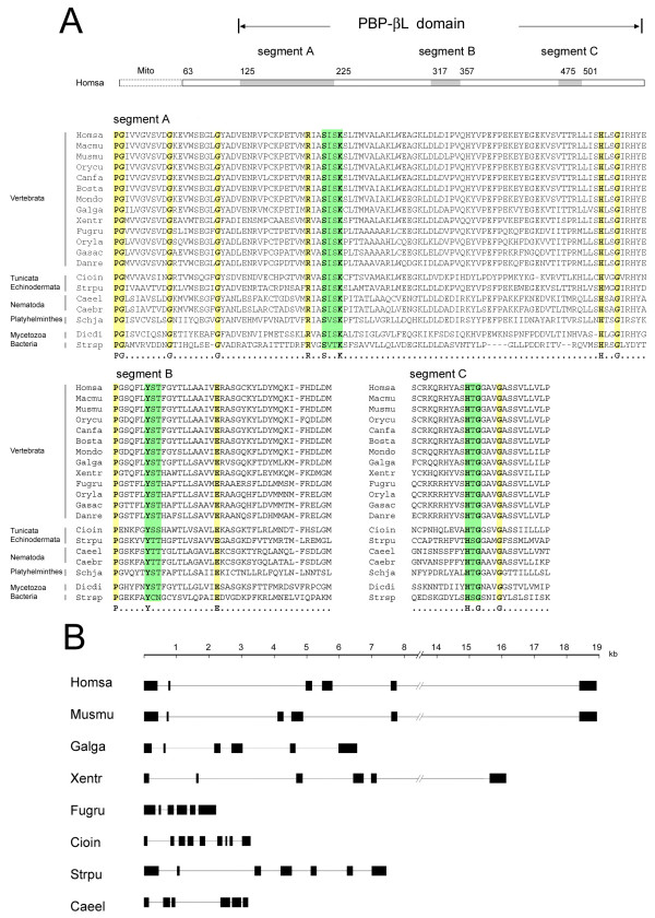
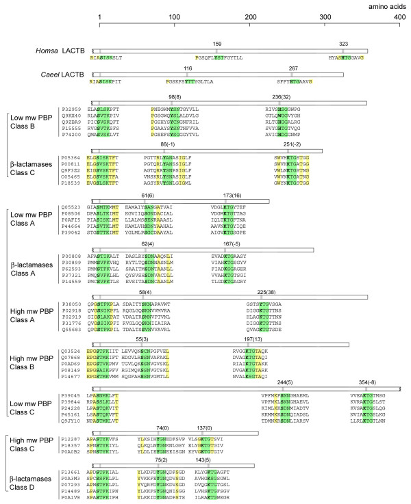
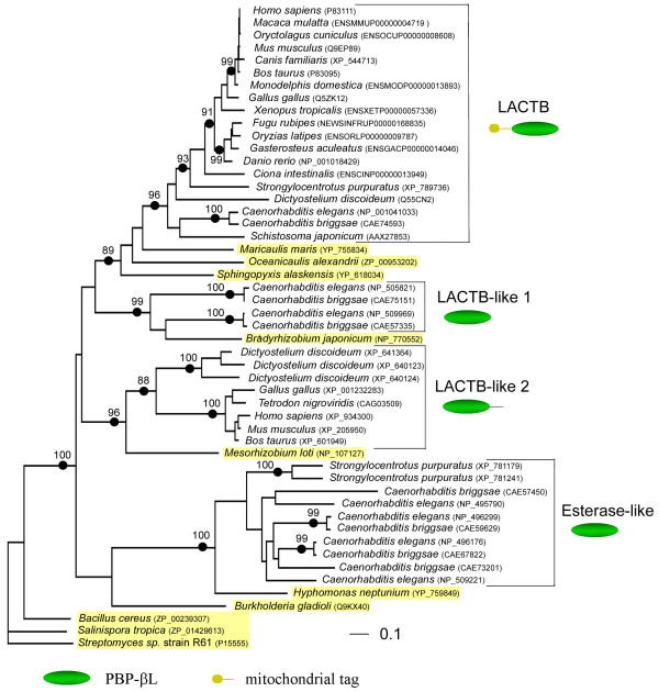
Results: Metazoan PBP-betaL homologs including LACTB share unique structural features with bacterial class B low molecular weight penicillin-binding proteins. The amino acid residues necessary for enzymatic activity in bacterial PBP-betaL proteins, including the catalytic serine residue, are conserved in all metazoan homologs. Phylogenetic analysis indicated that metazoan PBP-betaL homologs comprise four alloparalogus protein lineages that derive from alpha-proteobacteria.

Conclusion: While most components of the peptidoglycan synthesis machinery were dumped by early eukaryotes, a few PBP-betaL proteins were conserved and are found in metazoans including humans. Metazoan PBP-betaL homologs are active-site-serine enzymes that probably have distinct functions in the metabolic circuitry. We hypothesize that PBP-betaL proteins in the early eukaryotic cell enabled the degradation of peptidoglycan from ingested bacteria, thereby maximizing the yield of nutrients and streamlining the cell for effective phagocytotic feeding.


Figures

Figure 1
Figure 1
Amino acid alignment of catalytic site motifs and gene architecture of LACTB orthologs. (A) Schematic organization and alignment of the three PBP-βLs catalytic site motifs (highlighted in green) and their flanking regions in LACTB orthologs. The corresponding motifs in the Streptomyces sp. strain R61 D-alanyl-D-alanine carboxypeptidase [Swiss-Prot:P15555] are included. Abbreviations for species names, in order: Homsa, Homo sapiens (Swiss-Prot:P83111); Macmu, Macaca mulatta (Ensembl:ENSMMUP00000004719); Musmu, Mus musculus (Swiss-Prot:Q9EP89); Orycu, Oryctolagus cuniculus (Ensembl:ENSOCUP00000008608); Canfa, Canis familiaris (RefSeq:XP_544713); Bosta, Bos taurus (Swiss-Prot:P83095); Mondo, Monodelphis domestica (Ensembl:ENSMODP00000013893); Galga,Gallus gallus (Swiss-Prot:Q5ZK12); Xentr, Xenopus tropicalis (Ensembl:ENSXETG00000009720); Fugru, Fugu rubipes (Ensembl:SINFRUP00000138119); Oryla, Oryzia latipes (Ensembl:ENSORLP00000009787); Gasac, Gasterosteus aculeatus (Ensembl:ENSGACP00000014046); Danre, Danio rerio (RefSeq:NP_001018429); Strpu, Strongylocentrotus purpuratus (RefSeq:XP_789736); Cioin, Ciona intestinalis (Ensembl:ENSCING00000006798); Schja, Schistosoma japonicum (GenPept:AAX27853, AAX25200); Caeel, Caenorhabditis elegans (RefSeq:NP_001041033); Caebr, Caenorhabiditis briggsae (GenPept:CAE74593); Dicdi, Dictyostelium discoideum (Swiss-Prot:Q55CN2); Stesp, Streptomyces sp strain R61. The mitochondrial import sequence (Mito) is indicated. Amino acid conserved in all taxa are highlighted in yellow. (B) Organization of exons and introns in LACTB genes of representative metazoan taxa.
Figure 2
Figure 2
Schematic representation of the organization of the three catalytic site motifs in LACTB and the different PBP-βL classes. A set of founding members of each PBP-βL class (Additional file 1), classified according to Ghuysen 1997, and Massova and Mobashery 1998 [3,4], were used to calculate the median inter-motif distances in number of amino acid residues. Accession numbers refer to the Swiss-Prot database. The catalytic site motifs are highlighted in green and invariant amino acids are higlighted in yellow. Inter-motif distances were measured from the serine in the -SXXK-motif to the serine/lysine and lysine/histidine of the second and third catalytic site motif, respectively. Numbers within brackets is the largest difference from the median value within each class. PBP-βL classes forming separate clades [3] are marked with square brackets. Abbreviations: PBP, penicillin-binding protein.
Figure 3
Figure 3
Inferred phylogenetic tree of the PBP-βL domain of LACTB family proteins. An alignment encompassing 266 amino acids was analyzed by maximum likelihood as described in the Materials and Methods section. The log likelihood was -20487.28 and the Γ distribution shape parameter shape parameter used was 2.670. Bootstrap values for nodes supported by more than 85 replicates of 100 are shown. Bacterial proteins are highlighted in yellow.

References

    1. Macheboeuf P, Contreras-Martel C, Job V, Dideberg O, Dessen A. Penicillin Binding Proteins: key players in bacterial cell cycle and drug resistance processes. FEMS Microbiol Rev. 2006;30:673–691. doi: 10.1111/j.1574-6976.2006.00024.x. - DOI - PubMed
    1. Goffin C, Ghuysen J-M. Biochemistry and Comparative genomics of SxxK Superfamily Acyltransferases Offer a Clue to the Mycobacterial Paradox: Presence of Penicillin-Susceptible Target Proteins versus Lack of Efficiency of Penicillin as Therapeutic Agent. Microbiol Mol Biol Rev. 2002;66:702–738. doi: 10.1128/MMBR.66.4.702-738.2002. - DOI - PMC - PubMed
    1. Massova I, Mobashery S. Kinship and Diversification of Bacterial Penicillin-Binding Protein and β-Lactamases. Antimicrob Agents Chemother. 1998;42:1–17. - PMC - PubMed
    1. Ghuysen J-M. Penicillin-binding proteins. Wall peptidoglycan assembly and resistance to penicillin: facts, doubts and hopes. Int J Antimicrob Agents. 1997;8:45–60. doi: 10.1016/S0924-8579(96)00358-5. - DOI - PubMed
    1. Bush K, Jacoby GA, Medeiros AA. A Functional Classification Scheme for β-Lactamases and Its correlation with Molecular Structure. Antimicrob Agents Chemother. 1995;39:1211–1233. - PMC - PubMed

Publication types