Review
. 2008 Feb;20(1):41-7.
doi: 10.1016/j.ceb.2007.11.006. Epub 2008 Jan 15.

Cargo transport: molecular motors navigate a complex cytoskeleton

Affiliations
Review

Cargo transport: molecular motors navigate a complex cytoskeleton

Jennifer L Ross et al. Curr Opin Cell Biol. 2008 Feb.

Abstract

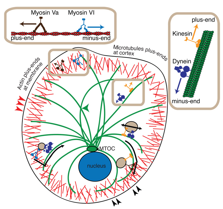
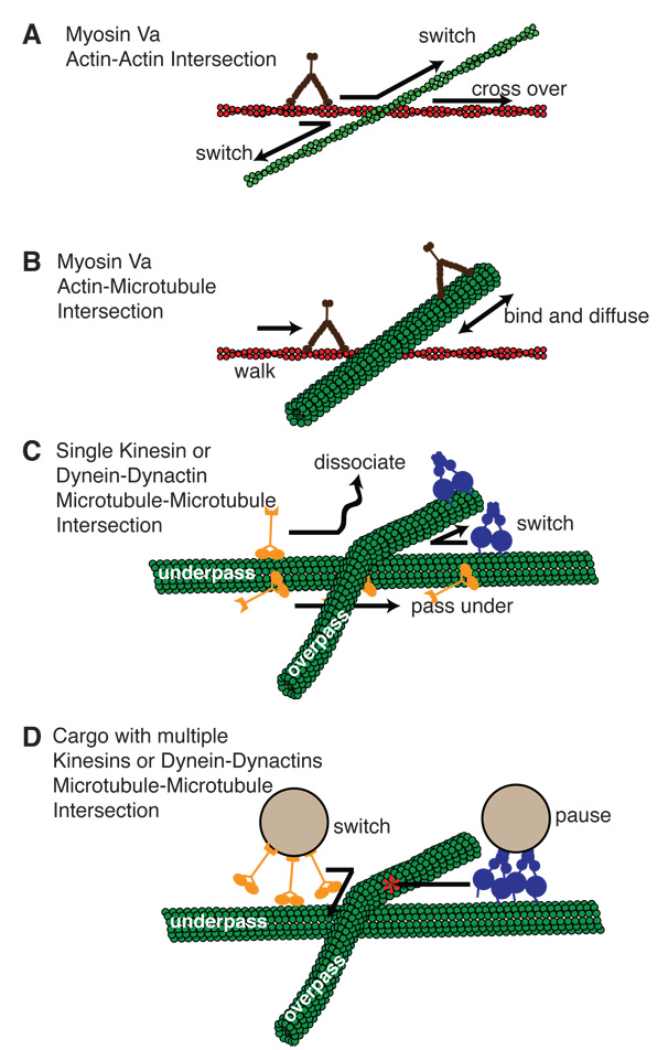
Intracellular cargo transport requires microtubule-based motors, kinesin and cytoplasmic dynein, and the actin-based myosin motors to maneuver through the challenges presented by the filamentous meshwork that comprises the cytoskeleton. Recent in vitro single molecule biophysical studies have begun to explore this process by characterizing what occurs as these tiny molecular motors happen upon an intersection between two cytoskeletal filaments. These studies, in combination with in vivo work, define the mechanism by which molecular motors exchange cargo while traveling between filamentous tracks and deliver it to its destination when going from the cell center to the periphery and back again.


Figures

Figure 1
Figure 1
Cartoon representation of motor proteins and vesicular cargo transport in the cell. Myosin family motors, myosin Va (dark brown) and myosin VI (light blue), walk along actin filaments (red) at the cortex. Myosin Va walks toward the F-actin plus-end, which is oriented towards the membrane. Myosin VI walks towards the minus-end of F-actin, toward the cell interior. Microtubule-based motors include the kinesin family motors (orange) and cytoplasmic dynein (violet). Kinesin motors walk to the plus-ends of microtubules (green), which are oriented toward the actin cortex. Dynein motors walk toward the minus-end of the microtubule, which is located at the microtubule-organizing center (MTOC, green) near the cell nucleus (blue). F-actin and microtubules cross at the cell cortex, as highlighted by black arrowheads (lower right). F-actin can cross itself in the cortex, highlighted by the red arrowheads (left). Microtubules can intersect other microtubules highlighted by the green arrowhead (center). Vesicular cargo (tan) can bind to myosin VI and dynein to switch from actin-based to microtubule-based motion while being transported into the cell interior (lower left). Vesicles can bind kinesin and myosin Va to switch from microtubule-based to actin-based motion in order to be transported to the cell cortex (lower right). Vesicles traveling on microtubules can experience a tug-of-war from kinesin and dynein simultaneously bound (right).
Figure 2
Figure 2
Schematic diagram of in vitro assays with intersecting cytoskeletal filaments. Intersections have been created between two F-actin filaments, an F-actin and a microtubule, and two microtubules. (A) For actin-actin intersections, recent studies have observed myosin Va switching most frequently to the crossing filament, but also passing over the intersecting F-actin (green). (B) The same study used the microtubule as an obstacle for Myosin Va stepping, but found myosin Va was able to bind and diffuse along microtubules. (C) An interesting feature of microtubule-microtubule intersections is that they have “underpass” and “overpass” tracks. Single kinesin are able to transgress the intersection on the underpass track by going under the overpass bridge. Dynein-dynactin was most likely to switch between microtubules. (D) Two studies of beads decorted with kinesin at microtubule-microtubule intersections have shown that kinesin coated-beads switch frequently at the intersection. Dynein-dynactin coated beads were found to pause at the intersection at high motor density, but pass or switch at low motor density.

References

    1. Chevalier-Larsen E, Holzbaur EL. Axonal transport and neurodegenerative disease. Biochim Biophys Acta. 2006;1762:1094–1108. - PubMed
    1. Lo Giudice M, Neri M, Falco M, Sturnio M, Calzolari E, Di Benedetto D, Fichera M. A missense mutation in the coiled-coil domain of the KIF5A gene and late-onset hereditary spastic paraplegia. Arch Neurol. 2006;63:284–287. - PubMed
    1. Takagishi Y, Murata Y. Myosin Va mutation in rats is an animal model for the human hereditary neurological disease, Griscelli syndrome type 1. Ann N Y Acad Sci. 2006;1086:66–80. - PubMed
    1. Lakadamyali M, Rust MJ, Babcock HP, Zhuang X. Visualizing infection of individual influenza viruses. Proc Natl Acad Sci U S A. 2003;100:9280–9285. - PMC - PubMed
    1. Kural C, Serpinskaya AS, Chou YH, Goldman RD, Gelfand VI, Selvin PR. Tracking melanosomes inside a cell to study molecular motors and their interaction. Proc Natl Acad Sci U S A. 2007;104:5378–5382. Melanosomes are localized with nm resolution inside cells and tracked as they move along microtubules and actin filaments. Single 8 nm steps powered by kinesin or dynein are observed for anterograde or retrograde movements along microtubules, with myosinV-powered 35 nm steps observed along actin filaments. Interestingly, a single melanasome was observed switching between microtubule and actin tracks as inferred from changes in direction that correlate with a change in step size

Publication types

Substances