

Abnormally increased CSF 3-Ortho-methyldopa (3-OMD) in untreated restless legs syndrome (RLS) patients indicates more severe disease and possibly abnormally increased dopamine synthesis
- PMID: 18226951
- PMCID: PMC2655320
- DOI: 10.1016/j.sleep.2007.11.012
Abnormally increased CSF 3-Ortho-methyldopa (3-OMD) in untreated restless legs syndrome (RLS) patients indicates more severe disease and possibly abnormally increased dopamine synthesis
Abstract
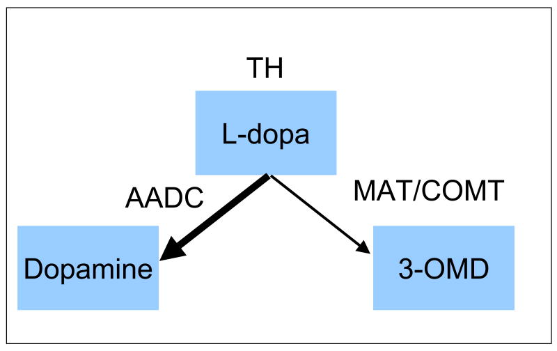
Background: Abnormally high CSF 3-OMD occurs frequently for RLS patients indicating either increased l-dopa synthesis, limitations in l-dopa decarboxylation or increased MAT/COMT activity, or some combination of these. Increased tyrosine hydroxylase activity was found on both the RLS autopsy and the rodent iron-deprivation model of RLS, suggesting increased DA synthesis in RLS. We, therefore, hypothesized elevated 3-OMD in RLS results from increased DA synthesis and that this should occur accordingly with increased HVA. It would then also reflect both the more severe iron insufficiency pathology of RLS and greater clinical severity, shown by the objective measure of PLMS/hr.
Methods: Patients off RLS medications and matched controls had lumbar punctures at either 10 a.m. or 10 p.m.; RLS patients were grouped by normal or abnormally high 3-OMD (>10 nmol/l).
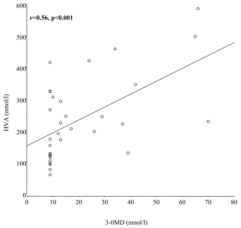
Results: Forty-nine RLS patients (30 high, 19 normal 3-OMD) and 36 age- and gender-matched controls, analyzed separately by time of CSF collection, did not significantly differ in age or gender. RLS patients with high 3-OMD had significantly higher CSF HVA, while those with normal 3-OMD had consistently lower CSF HVA than controls. CSF ferritin was consistently lower compared to controls for the high 3-OMD but not the normal 3-OMD RLS patients. The PLMS/hr was significantly higher for RLS patients with high compared to normal 3-OMD, indicating high 3-OMD patients had more severe RLS.
Conclusions: Abnormal elevation in 3-OMD for RLS patients may reflect increased dopamine synthesis for more severe but perhaps not mild RLS. These differences in the putative dopamine pathology of RLS may indicate different phases or expression of RLS biology or different underlying disease processes.
Figures
References
-
- Turjanski N, Lees AJ, Brooks DJ. Striatal dopaminergic function in restless legs syndrome: 18F-dopa and 11C-raclopride PET studies. Neurology. 1999;52(5):932–7. - PubMed
-
- Michaud M, Soucy JP, Chabli A, Lavigne G, Montplaisir J. SPECT imaging of striatal pre- and postsynaptic dopaminergic status in restless legs syndrome with periodic leg movements in sleep. J Neurol. 2002 Feb;249(2):164–70. - PubMed
-
- Ruottinen HM, Partinen M, Hublin C, Bergman J, Haaparanta M, Solin O, et al. An FDOPA PET study in patients with periodic limb movement disorder and restless legs syndrome. Neurology. 2000;54(2):502–4. - PubMed
-
- Cervenka S, Palhagen SE, Comley RA, Panagiotidis G, Cselenyi Z, Matthews JC, et al. Support for dopaminergic hypoactivity in restless legs syndrome: a PET study on D2-receptor binding. Brain. 2006 Jul 1; - PubMed
-
- Tribl GG, Asenbaum S, Klosch G, Mayer K, Bonelli RM, Auff E, et al. Normal IPT and IBZM SPECT in drug naive and levodopa-treated idiopathic restless legs syndrome (letter to editor) Neurology. 2002 Aug 27;59(4):649–50. - PubMed
MeSH terms
Substances
Grants and funding
LinkOut - more resources
Full Text Sources
Medical
Miscellaneous
