. 2008 Apr 4;283(14):9341-9.
doi: 10.1074/jbc.M707807200. Epub 2008 Jan 27.

Anandamide biosynthesis catalyzed by the phosphodiesterase GDE1 and detection of glycerophospho-N-acyl ethanolamine precursors in mouse brain

Affiliations

Anandamide biosynthesis catalyzed by the phosphodiesterase GDE1 and detection of glycerophospho-N-acyl ethanolamine precursors in mouse brain

Gabriel M Simon et al. J Biol Chem. .

Abstract

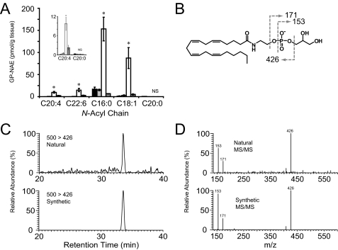
Anandamide (AEA) is an endogenous ligand of cannabinoid receptors and a well characterized mediator of many physiological processes including inflammation, pain, and appetite. The biosynthetic pathway(s) for anandamide and its N-acyl ethanolamine (NAE) congeners remain enigmatic. Previously, we proposed an enzymatic route for producing NAEs that involves the double-O-deacylation of N-acyl phosphatidylethanolamines (NAPEs) by alpha/beta-hydrolase 4 (ABDH4 or Abh4) to form glycerophospho (GP)-NAEs, followed by conversion of these intermediates to NAEs by an unidentified phosphodiesterase. Here, we report the detection and measurement of GP-NAEs, including the anandamide precursor glycerophospho-N-arachidonoylethanolamine (GP-NArE), as endogenous constituents of mouse brain tissue. Inhibition of the phosphodiesterase-mediated degradation of GP-NAEs ex vivo resulted in a striking accumulation of these lipids in brain extracts, suggesting a rapid endogenous flux through this pathway. Furthermore, we identify the glycerophosphodiesterase GDE1, also known as MIR16, as a broadly expressed membrane enzyme with robust GP-NAE phosphodiesterase activity. Together, these data provide evidence for a multistep pathway for the production of anandamide in the nervous system by the sequential actions of Abh4 and GDE1.


Figures

FIGURE 1.
FIGURE 1.
Proposed biosynthetic pathway for NAEs including AEA. Both O-acyl chains are removed from N-arachidonoyl phosphatidylethanolamine (NArPE) by Abh4 yielding GP-NArE. The phosphodiester bond of GP-NArE is then hydrolyzed by a phosphodiesterase (identified herein as GDE1) to release free anandamide and glycerol 3-phosphate.
FIGURE 2.
FIGURE 2.
Fragmentation and chromatography of endogenous GP-NAEs in mouse brain via LC-MS/MS. Fragmentation and chromatographic behavior are shown for endogenous N-palmitoyl (A-C) and N-oleoyl (D-F) GP-NAEs. Three major fragment ions are produced, a diagnostic NAE-phosphate fragment (378 and 404 for palmitoyl- and oleoyl-GP-NAEs, respectively) and two fragments corresponding to glycerol 3-phosphate and its dehydrate at 171 and 153, respectively (A and D). B and E, representative multiple reaction monitoring chromatographs of synthetic and endogenous GP-NAEs. C and F, tandem MS/MS fragmentation spectra of synthetic and endogenous GP-NAEs.
FIGURE 3.
FIGURE 3.
Ex vivo accumulation of GP-NAEs in brain tissue via an MAFP-sensitive and EDTA-dependent enzymatic pathway. A, GP-NAE levels in brain tissue homogenized in buffer (50 mm Tris, pH 8.0) alone (black and light gray bars), buffer with 10 mm EDTA (open bars) or 10 mm EDTA and 20 μm MAFP (dark gray bars). Homogenates were incubated for 0 h (black bars) or 4 h (all other bars) and then subjected to organic extraction and LC-MS/MS analysis. The inset shows a magnification of low abundance GP-NAEs. Student's t test compared EDTA-treated to samples treated with EDTA and MAFP; asterisk indicates p < 0.05; NS, not significant (n = 4 per group). B, fragmentation of N-arachidonoyl-GP-NAE produces a diagnostic NAE-phosphate fragment (m/z 426) and two fragments corresponding to glycerol 3-phosphate and its dehydrate at 171 and 153, respectively. Representative chromatographs (C) and tandem MS fragmentation (D) are shown for synthetic GP-NArE and natural GP-NArE, which accumulates in the presence of EDTA.
FIGURE 4.
FIGURE 4.
Expression and GP-NAE phosphodiesterase activity of GDE domain containing enzymes. A, Myc-tagged GDE-domain containing enzymes were overexpressed in COS-7 cells. Membrane fractions were treated with PNGase F and visualized by SDS-PAGE and Western blotting with an anti-Myc antibody (Cell Signaling). B, GP-NAE phosphodiesterase activity of membrane fractions of COS-7 cells expressing GDE domain-containing proteins was measured by monitoring release of N-C16:0 NAE via LC-MS with 100 μm N-C16:0 GP-NAE as substrate.
FIGURE 5.
FIGURE 5.
GDE1 and GP-NAE phosphodiesterase activity reside in the membrane. A, Western blot of mouse brain GDE1 showing that it resides exclusively in the membrane fraction and is a glycoprotein as evidenced by a ∼5-kDa shift in migration upon treatment with PNGase F. B, GP-NAE phosphodiesterase activity resides exclusively in the membrane fraction of mouse brain.
FIGURE 6.
FIGURE 6.
Mouse brain GP-NAE phosphodiesterase and GDE1 display similar biochemical properties. A, pH rate profile of brain membrane GP-NAE phosphodiesterase (open diamonds) and membranes from COS-7 cells transfected with GDE1 (filled squares) showing equivalent pH optima. Assays were performed in buffer containing 50 mm Tris, 50 mm HEPES, and 50 mm glycine and adjusted to the desired pH with HCl/NaOH. B, mouse brain GP-NAE phosphodiesterase and GDE1 display similar cation sensitivity profiles. Mouse brain membranes (open bars) and GDE1-transfected COS-7 cell membranes (filled bars) were treated with 2 mm CaCl2, ZnCl2, MgCl2, or EDTA. Assays were performed with 100 μm [1′-14C]N-C16:0 GP-NAE substrate, where the release of [1′-14C]N-C16:0 NAE was measured by thin layer radiochromatography.
FIGURE 7.
FIGURE 7.
Tissue distribution of GP-NAE phosphodiesterase activity and GDE1. A, GP-NAE phosphodiesterase activity in membrane fractions of mouse tissues. Assays were performed with 100 μm [1′-14C]N-C16:0 GP-NAE substrate, where the release of [1′-14C]N-C16:0 NAE was measured by thin layer radiochromatography. B, Western blot of GDE1 expression in membrane fractions of mouse tissues following treatment with PNGase F showing strong correlation with GP-NAE phosphodiesterase activity. C, reverse transcription-PCR analysis of GDE1 mRNA expression correlates with GDE1 protein levels and GP-NAE phosphodiesterase activity. The ubiquitously expressed enzyme glyceraldehyde-3-phosphate dehydrogenase (GAPDH) was used as a control to confirm integrity of tissue cDNA. Br, brain; Spc, spinal cord; Ht, heart; Lv, liver; Kd, kidney; Spln, spleen; Ts, testis.
FIGURE 8.
FIGURE 8.
N-Acyl chain selectivity of GDE1. Membrane fractions from GDE1-transfected COS-7 cells were tested for activity against a panel of GP-NAE substrates bearing saturated (C16:0, C20:0), monounsaturated (C18:1), and polyunsaturated (C20:4, C22:6) N-acyl chains. Assays were performed with 100 μm of each substrate using LC-MS to measure release of NAE.

References

    1. Kuehl, F. A., Jacob, T. A., Ganley, O. H., Ormond, R. E., and Meisinger, M. A. P. (1957) J. Am. Chem. Soc. 79 5577-5578
    1. Bachur, N. R., Masek, K., Melmon, K. L., and Udenfriend, S. (1965) J. Biol. Chem. 240 1019-1024 - PubMed
    1. Natarajan, V., Reddy, P. V., Schmid, P. C., and Schmid, H. H. (1982) Biochim. Biophys. Acta 712 342-355 - PubMed
    1. Devane, W. A., Hanus, L., Breuer, A., Pertwee, R. G., Stevenson, L. A., Griffin, G., Gibson, D., Mandelbaum, A., Etinger, A., and Mechoulam, R. (1992) Science 258 1946-1949 - PubMed
    1. Cravatt, B. F., Giang, D. K., Mayfield, S. P., Boger, D. L., Lerner, R. A., and Gilula, N. B. (1996) Nature 384 83-87 - PubMed

Publication types