. 2008 Mar 28;283(13):8301-9.
doi: 10.1074/jbc.M706730200. Epub 2008 Jan 27.

Regulation of myoblast motility and fusion by the CXCR4-associated sialomucin, CD164

Affiliations

Regulation of myoblast motility and fusion by the CXCR4-associated sialomucin, CD164

Gyu-Un Bae et al. J Biol Chem. .

Abstract

Myoblast fusion is fundamental to the development and regeneration of skeletal muscle. To fuse, myoblasts undergo cell-cell recognition and adhesion and merger of membranes between apposing cells. Cell migration must occur in advance of these events to bring myoblasts into proximity, but the factors that regulate myoblast motility are not fully understood. CD164 is a cell surface sialomucin that is targeted to endosomes and lysosomes via its intracellular region. In hematopoietic progenitor cells, CD164 forms complexes with the motility-stimulating chemokine receptor, CXCR4, in response to the CXCR4 ligand, CXCL12/SDF-1 (Forde, S., Tye, B. J., Newey, S. E., Roubelakis, M., Smythe, J., McGuckin, C. P., Pettengell, R., and Watt, S. M. (2007) Blood 109, 1825-1833). We have previously shown that CD164 stimulates myotube formation in vitro. We report here that CD164 is associated with CXCR4 in C2C12 myoblasts. Cells in which CD164 levels are increased or decreased via overexpression or RNA interference-mediated knockdown, respectively, show enhanced or reduced myotube formation and cell migration, the latter both basally and in response to CXCL12/SDF-1. Furthermore, expression of CD164 cytoplasmic tail mutants that alter the endosome/lysosome targeting sequence and, consequently, the subcellular localization in myoblasts, reveals a similar correlation between cell motility and myotube formation. Finally, Cd164 mRNA is expressed in the dorsal somite (the early myogenic compartment of the mouse embryo) and in premuscle masses. Taken together, these results suggest that CD164 is a regulator of myoblast motility and that this property contributes to its ability to promote myoblast fusion into myotubes.


Figures

FIGURE 1.
FIGURE 1.
Whole mount RNA in situ hybridization analysis of Cd164 expression in mouse embryos. A, side view of E9.5 embryo. B, axial view of E9.5 embryo. C, side view of E10.5 embryo. The white arrows indicate dorsal somites.
FIGURE 2.
FIGURE 2.
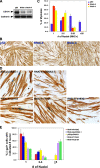
Expression of GFP-CD164 enhances myotube formation by C2C12 cells. A, 293T cells were transiently transfected with the indicated expression vectors encoding wild-type or GFP-tagged CD164 or the appropriate control vectors, and cell lysates were analyzed by Western blotting techniques with antibodies to CD164 (left panel) or GFP (right panel). B, C2C12 cells were stably transfected with pcDNA3.1 encoding GFP-CD164 (+) or, as a control, pcDNA3.1 itself (–), and cell lysates were analyzed by Western blotting techniques with antibodies to CD164 or, as a loading control, cadherin. C and D, C2C12 cell lines indicated in B (designated + CD164 and control, respectively) were cultured in DM, fixed and stained with an antibody to MHC (C), and then quantified for the percentage of MHC+ nuclei present in myotubes that contained the indicated number of nuclei (D). Values represent means of triplicate determinations ± S.D.
FIGURE 3.
FIGURE 3.
GFP-CD164 cytoplasmic tail mutants have distinct subcellular locations in C2C12 myoblasts. A, sequence of the intracellular region of CD164 and of three mutants. WT, wild type. B and C, confocal micrographs of C2C12 myoblasts (B) and nascent myotubes (C) expressing GFP-CD164 (CD164) or the indicated GFP-CD164 mutants. The myoblast cultures were also stained with an antibody that recognizes classical cadherins, including two that are expressed in C2C12 cells, N- and M-cadherin. D, biochemical fractionation of C2C12 myoblasts expressing GFP-CD164 (CD164) or the indicated GFP-CD164 mutants into vesicle and membrane preparations. The vesicle and membrane fractions were analyzed by Western blotting with antibodies to the indicated proteins. Antibodies against the early endosomal marker EEA1 and classical cadherins were used to demonstrate the purity of the vesicle and membrane fractions, respectively. A portion of the total cell lysate was also probed with antibodies to GFP to document approximately equivalent expression levels of each CD164 derivative. E, the pcDNA3.1 control vector lanes from D were probed with antibody against CD164 to reveal relative vesicle and membrane distribution of endogenous CD164 in C2C12 cells. Note that the EEA1 and cadherin signals in D are therefore the appropriate controls (E).
FIGURE 4.
FIGURE 4.
Myotube formation by C2C12 cells that stably express GFP-CD164 cytoplasmic tail mutants. A, Western blot analysis of expression of GFP-CD164 and the indicated mutants. Cell lysates were analyzed by Western blotting techniques with antibodies to GFP or, as a loading control, cadherin. B and C, C2C12 cell lines indicated in A were cultured in DM, fixed and stained with an antibody to MHC (B), and then quantified for the percentage of MHC+ nuclei present in myotubes that contained the indicated number of nuclei (C). Values represent means of triplicate determinations ± S.D.
FIGURE 5.
FIGURE 5.
Reduction of C2C12 myotube formation by RNAi against Cd164 and rescue by GFP-CD164 constructs. A, Western blot analysis of CD164 levels by two independent RNAi sequences. Cell lysates were analyzed by Western blotting techniques with antibodies to CD164 or, as a loading control, cadherin. pSil, pSilencer. B and C, C2C12 cell lines indicated in A were cultured in DM, fixed and stained with an antibody to MHC (B), and then quantified for the percentage of MHC+ nuclei present in myotubes that contained the indicated number of nuclei (C). Values represent means of triplicate determinations ± S.D. D and E, C2C12 cells were transiently transfected with pSilencer or pSilencer driving expression of Cd164 RNAi-4 plus pcDNA3.1 or pcDNA3.1 driving expression of GFP-CD164 or the indicated cytoplasmic tail mutants in each case plus an expression vector for nuclear localized β-galactosidase (β-gal). Cultures were fixed and double-stained for MHC and β-galactosidase activity (D) and myotube formation by transfectants, which was quantified as the percentage of β-galactosidase+ cells with the indicated number of nuclei (E). Values represent means of triplicate determinations ± S.D. Note that the level of myotube formation by the control cultures in panels B and C and panels D and E (pSil cells and pSil/pcDNA3.1 cells, respectively) is different (∼18% mononucleate MHC+ pSil cells in B and C and ∼55% mononucleate MHC+ pSil/pcDNA3.1 cells in D and E). These conditions were selected so as to permit easy visualization of diminished myotube formation by Cd164 RNAi in (B) and (C) and either enhanced or diminished myotube formation by the various CD164 mutants in (D) and (E). See “Results” for further details.
FIGURE 6.
FIGURE 6.
Co-immunoprecipitation of CD164 and CXCR4. A, C2C12 cells were analyzed for association of CD164 and CXCR4 through a time course of differentiation. Cells were harvested from proliferating cultures in growth medium (G), from cultures at ∼80% confluence at the time of transfer to DM (D0), and from cultures in DM for 1, 2, or 3 days (D1, D2, and D3, respectively). Cell lysates were immunoprecipitated (IP) with the indicated antibodies and then analyzed by Western blotting (WB) techniques with the reciprocal antibody. Straight cell lysates were also analyzed by Western blotting with the indicated antibodies. MHC reveals the progression of differentiation, and cadherin serves as a loading control. B, RT-PCR analysis of CXCL12 mRNA expression and, as a control, GAPDH mRNA expression in C2C12 cells through a time course of differentiation.
FIGURE 7.
FIGURE 7.
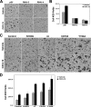
Motility of C2C12 cells expressing RNAi against Cd164, GFP-CD164, or GFP-CD164 cytoplasmic tail mutants. A and B, C2C12 cells expressing the indicated RNAi sequences against Cd164 or control vector (pSilencer (pSil)) were analyzed for migration in Boyden chambers in response to control medium or medium containing CXCL12. Cells that had migrated were photographed (A) and quantified (B). Values represent means of triplicate determinations ± S.D. C and D, C2C12 cells expressing the indicated GFP-CD164 variant or control vector (control) were analyzed for migration in Boyden chambers in response to control medium or medium containing CXCL12. Cells that had migrated were photographed (C) and quantified (D). Values represent means of triplicate determinations ± S.D.

References

    1. Tajbakhsh, S., and Buckingham, M. (2000) Curr. Top. Dev. Biol. 48 225–268 - PubMed
    1. Horsley, V., and Pavlath, G. K. (2004) Cells Tissues Organs 176 67–78 - PubMed
    1. Perry, R. L. S., and Rudnicki, M. A. (2000) Front. Biosci. 5 d750–767 - PubMed
    1. Tapscott, S. J. (2005) Development (Camb.) 132 2685–2695 - PubMed
    1. Krauss, R. S., Cole, F., Gaio, U., Takaesu, G., Zhang, W., and Kang, J. S. (2005) J. Cell Sci. 118 2355–2362 - PubMed

Publication types

MeSH terms