. 2008 Mar 28;283(13):8462-8.
doi: 10.1074/jbc.M709572200. Epub 2008 Jan 28.

Regulation of nuclear lamin polymerization by importin alpha

Affiliations

Regulation of nuclear lamin polymerization by importin alpha

Stephen A Adam et al. J Biol Chem. .

Abstract

Nuclear lamins are integral components of the nuclear envelope and are important for the regulation of many aspects of nuclear function, including gene transcription and DNA replication. During interphase, the lamins form an intranuclear intermediate filament network that must be disassembled and reassembled when cells divide. Little is known about factors regulating this assembly/disassembly cycle. Using in vitro nuclear assembly and lamin assembly assays, we have identified a role for the nuclear transport factor importin alpha in the regulation of lamin assembly. Exogenous importin alpha inhibited nuclear lamin assembly in Xenopus interphase egg nuclear assembly assays. Fractionation of the egg extract used for nuclear assembly identified a high molecular weight complex containing the major egg lamin, XLB3, importin alpha, and importin beta. This complex could be dissociated by RanGTP or a competing nuclear localization sequence, indicating that lamin assembly is Ran- and importin alpha-dependent in the egg extract. We show that the addition of importin alpha to purified lamin B3 prevents the assembly of lamins in solution. Lamin assembly assays show that importin alpha prevents the self-association of lamins required to assemble lamin filaments into the typical paracrystals formed in vitro. These results suggest a role for importin alpha in regulating lamin assembly and possibly modulating the interactions of lamins with lamin-binding proteins.


Figures

FIGURE 1.
FIGURE 1.
Inhibition of nuclear assembly by importin α. Interphase egg extracts were pre-incubated with 10 μm MBP (a–c) or with 10 μm importin α (Impα; d–f) for 30 min prior to the introduction of demembranated sperm chromatin. After a 75-min incubation at 23 °C, the assembled nuclei were centrifuged onto poly-l-lysine-coated coverslips, fixed, and processed for immunofluorescence. XLB3 was detected with a polyclonal antiserum to XLB3, and nucleoporins (Nup) were detected with MAb414. Images were collected on a Zeiss Axiovision microscope. Optimal exposure times were determined for the control assembly reactions in a–c and used to image assembly reactions with importin α in d and e. In f, the brightness and contrast were adjusted to the same levels as in a–c to more clearly show the protein localization. All images were processed using Zeiss AxiovisionLE version 4.2 software. The scale bar on each panel = 10 μm.
FIGURE 2.
FIGURE 2.
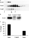
XLB3 is present in a large complex with importin α. A, demembranated egg cytosol was fractionated by gel filtration chromatography. Duplicate immunoblots were probed with antibodies to XLB3, importin α (Impα), and importin β (Impβ). The performance of the Superose 6 column was determined by separation of purified standard proteins in the same buffer as the egg cytosol. v, void volume; tg, thyroglobulin (Mr 669,000); f, ferritin (Mr 440,000). B, aliquots of the peak fraction of XLB3 were immunoadsorbed with immobilized antibodies to importin α. Pre-immune serum was used as the control. The bound proteins were analyzed by immunoblotting with antibodies to XLB3. The same amount of XLB3 added to the immunoadsorption was loaded in the first lane (Input). C, two independent immunadsorptions were performed as in B, and the amount of XLB3 adsorbed was quantified as described under “Experimental Procedures.” The error bars indicate average deviation.
FIGURE 3.
FIGURE 3.
Release of XLB3 from importin complex by RanGTP. The pooled XLB3 peak fractions from the gel filtration chromatography in Fig. 2 were incubated with 5 μm concentrations of the indicated proteins to release XLB3. After incubation, the soluble and insoluble XLB3 were separated by centrifugation as described under “Experimental Procedures.” The soluble (S) and pelleted (P) fractions were separated by SDS-PAGE and immunoblotted with antibodies to XLB3. Band intensity was determined with a Kodak 440CF Image Station.
FIGURE 4.
FIGURE 4.
Importin α prevents XLB3 aggregation in vitro. Bacterially expressed XLB3 or XLB3ΔNLS was dialyzed under buffer conditions that promote aggregation of lamins. Importin α (Impα) or purified MBP was added at a 5-fold molar excess over the concentration of XLB3 to inhibit aggregation. After incubation, the mixture was separated into soluble and insoluble (pellet) fractions by centrifugation. The soluble and pellet fractions were separated by SDS-PAGE, and the amount of XLB3 in each fraction was determined by Coomassie Brilliant Blue G-250 staining and quantification using a Kodak Image Station as described under “Experimental Procedures.” The amount of pelletable XLB3 is expressed as a percentage of the total XLB3 added. The error bars indicate average deviation of data from duplicate experiments. WT, wild-type.
FIGURE 5.
FIGURE 5.
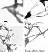
Importin α inhibits formation of XLB3 paracrystals. Paracrystals of XLB3 were formed as described under “Experimental Procedures” and visualized by electron microscopy after negative staining. Importin α (Impα) was added in a 5-fold molar excess to inhibit paracrystal formation. For all micrographs, the magnification is ×25,000, and the scale bar = 200 nm. WT, wild-type.

References

    1. Burke, B. (1990) Curr. Opin. Cell Biol. 2 514-520 - PubMed
    1. Mattout, A., Dechat, T., Adam, S. A., Goldman, R. D., and Gruenbaum, Y. (2006) Curr. Opin. Cell Biol. 18 335-341 - PubMed
    1. Dorner, D., Gotzmann, J., and Foisner, R. (2007) FEBS J. 274 1362-1373 - PubMed
    1. Shumaker, D. K., Kuczmarski, E. R., and Goldman, R. D. (2003) Curr. Opin. Cell Biol. 15 358-366 - PubMed
    1. Nakajima, N., and Abe, K. (1995) FEBS Lett. 365 108-114 - PubMed

Publication types

LinkOut - more resources