. 2008 Apr 4;283(14):9435-43.
doi: 10.1074/jbc.M709135200. Epub 2008 Jan 28.

Structural basis for substrate fatty acyl chain specificity: crystal structure of human very-long-chain acyl-CoA dehydrogenase

Affiliations

Structural basis for substrate fatty acyl chain specificity: crystal structure of human very-long-chain acyl-CoA dehydrogenase

Ryan P McAndrew et al. J Biol Chem. .

Abstract

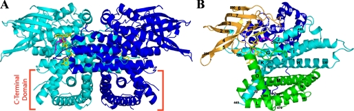
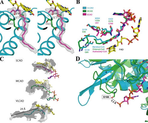
Very-long-chain acyl-CoA dehydrogenase (VLCAD) is a member of the family of acyl-CoA dehydrogenases (ACADs). Unlike the other ACADs, which are soluble homotetramers, VLCAD is a homodimer associated with the mitochondrial membrane. VLCAD also possesses an additional 180 residues in the C terminus that are not present in the other ACADs. We have determined the crystal structure of VLCAD complexed with myristoyl-CoA, obtained by co-crystallization, to 1.91-A resolution. The overall fold of the N-terminal approximately 400 residues of VLCAD is similar to that of the soluble ACADs including medium-chain acyl-CoA dehydrogenase (MCAD). The novel C-terminal domain forms an alpha-helical bundle that is positioned perpendicular to the two N-terminal helical domains. The fatty acyl moiety of the bound substrate/product is deeply imbedded inside the protein; however, the adenosine pyrophosphate portion of the C14-CoA ligand is disordered because of partial hydrolysis of the thioester bond and high mobility of the CoA moiety. The location of Glu-422 with respect to the C2-C3 of the bound ligand and FAD confirms Glu-422 to be the catalytic base. In MCAD, Gln-95 and Glu-99 form the base of the substrate binding cavity. In VLCAD, these residues are glycines (Gly-175 and Gly-178), allowing the binding channel to extend for an additional 12A and permitting substrate acyl chain lengths as long as 24 carbons to bind. VLCAD deficiency is among the more common defects of mitochondrial beta-oxidation and, if left undiagnosed, can be fatal. This structure allows us to gain insight into how a variant VLCAD genotype results in a clinical phenotype.


Figures

FIGURE 1.
FIGURE 1.
A, ribbon diagram of the overall fold of the human VLCAD dimer. The monomers are represented in cyan and blue. The FADs are shown in yellow, and a partial model of C14-CoA is shown in magenta. The C-terminal domain is marked near the bottom of the figure. B, overall polypeptide fold of a VLCAD monomer showing the N-terminal α-dom1 (blue), the β-sheet domain (gold), α-dom2 (cyan), and the C-terminal α-dom3 (green). The FAD cofactor and the partially hydrolyzed model of substrate/product (C14-CoA) are shown with sticks in yellow and pink, respectively. α-Helices are labeled alphabetically, and β-strands are numbered consecutively from the N to C terminus. The numbers in a smaller font are residue numbers. Unless otherwise noted, all figures were generated using Pymol (52).
FIGURE 2.
FIGURE 2.
Superposition of: A, VLCAD dimer (blue and cyan) on an MCAD dimer (red and orange; PDB 3MDE); B, VLCAD monomer (blue) on an MCAD tetramer (shades of brown and red; PDB 3MDE) with a close-up of the overlay of the C-terminal domain with the other monomers; C, VLCAD monomer (blue) on an ACO monomer (magenta; PDB 2DDH).
FIGURE 3.
FIGURE 3.
A, stereo diagram of a 2Fo-Fc electron density map (1.0σ) fitted with a partial model of C14-CoA. The catalytic base, Glu-422 (green), is located in a position analogous to the catalytic residues of MCAD and SCAD. As with MCAD and SCAD, substrate position is stabilized by hydrogen bonds between the thioester carbonyl and both the amide nitrogen of the catalytic glutamate (2.7 Å) and the ribityl 2′-hydroxyl of FAD (2.9 Å). Helices are labeled. Part of the pantothenic acid and adenosine pyrophosphate moiety of CoA are not visible. The thioester sulfur is shown as green for clarity. B, overlay of the binding site residues of VLCAD (blue), MCAD (green; ID: 3MDE), and SCAD (magenta; ID: 1JQI). Gln-95 and Glu-99 in MCAD form the bottom of its binding cavity. In VLCAD, the corresponding residues are glycines (Gly-135 and Gly-138). This effectively extends the cavity and allows for much longer substrates to bind. C, surface renderings (inside, light gray; outside, dark gray) of the binding cavities of SCAD, MCAD, and VLCAD illustrate the basis of their substrate chain length specificities. The cavity depths of SCAD, MCAD, and VLCAD, measured from the substrate thioester carbonyls (shown for VLCAD), are 8, 12, and 24 Å. Only a partial model of the CoA moiety of C14-CoA is shown. D, overlay of the substrate binding sites of VLCAD (cyan) and MCAD (green; ID: 3MDE). In MCAD, residue Ser-166 on the loop between β-strands 4 and 5 hydrogen bonds with the 3′-phosphate on the CoA moiety of octanoyl-CoA. In VLCAD, the β-sheet formed by strands 4 and 5 extends much further away from the substrate binding site, thereby precluding an analogous hydrogen bond, and resulting in a wider opening of the substrate binding cavity.
FIGURE 4.
FIGURE 4.
Putative membrane binding site of VLCAD. A, an electrostatic representation (top) and a ribbon representation of the membrane-binding surface are shown (90° rotation from Fig. 1 along the x-axis). B, a helix model built from residues 441-481, which include the disordered residues. The helix shows a periodicity that is strikingly amphipathic in nature and may be capable of interacting with the mitochondrial membrane. C, putative VLCAD membrane orientation. It is proposed that residues 445-479 are responsible for anchoring VLCAD to the membrane in the orientation pictured. D, magnification and stereo view of the rectangular region of Fig. 4C. The dotted purple line represents the disordered region.
FIGURE 5.
FIGURE 5.
A, site of the R429W clinical mutation. Arg-429 (cyan) on helix K forms a salt bridge with Glu-384 (green) of helix I, which positions the catalytic glutamate, Glu-422 (orange). Truncated C14-CoA and FAD are depicted as magenta and yellow, respectively. B, site of the R416H mutation. Arg-416 forms a salt bridge with Asp-391 and a hydrogen bond with Gln-395 on the other monomer, thus stabilizing the dimer. Residues on the opposing monomer are labeled with an asterisk.

References

    1. Beinert, H. (1963) in The Enzymes. (Boyer, P. D., Lardy, H., and Myrback, K., eds) Vol. 7, pp. 447-476, Academic Press, New York
    1. Aoyama, T., Souri, M., Ushikubo, S., Kamijo, T., Yamaguchi, S., Kelley, R. I., Rhead, W. J., Uetake, K., Tanaka, K., and Hashimoto, T. (1995) J. Clin. Investig. 95 2465-2473 - PMC - PubMed
    1. Ikeda, Y., Okamura-Ikeda, K., and Tanaka, K. (1985) J. Biol. Chem. 260 1311-1325 - PubMed
    1. Izai, K., Uchida, Y., Orii, T., Yamamoto, S., and Hashimoto, T. (1992) J. Biol. Chem. 267 1027-1033 - PubMed
    1. Ensenauer, R., He, M., Willard, J. M., Goetzman, E. S., Corydon, T. J., Vandahl, B. B., Mohsen, A. W., Isaya, G., and Vockley, J. (2005) J. Biol. Chem. 280 32309-32316 - PubMed

Publication types

MeSH terms

Associated data