. 2008 Mar 28;283(13):8545-54.
doi: 10.1074/jbc.M709020200. Epub 2008 Jan 28.

Identification of a GH110 subfamily of alpha 1,3-galactosidases: novel enzymes for removal of the alpha 3Gal xenotransplantation antigen

Affiliations

Identification of a GH110 subfamily of alpha 1,3-galactosidases: novel enzymes for removal of the alpha 3Gal xenotransplantation antigen

Qiyong P Liu et al. J Biol Chem. .

Abstract

In search of alpha-galactosidases with improved kinetic properties for removal of the immunodominant alpha1,3-linked galactose residues of blood group B antigens, we recently identified a novel prokaryotic family of alpha-galactosidases (CAZy GH110) with highly restricted substrate specificity and neutral pH optimum (Liu, Q. P., Sulzenbacher, G., Yuan, H., Bennett, E. P., Pietz, G., Saunders, K., Spence, J., Nudelman, E., Levery, S. B., White, T., Neveu, J. M., Lane, W. S., Bourne, Y., Olsson, M. L., Henrissat, B., and Clausen, H. (2007) Nat. Biotechnol. 25, 454-464). One member of this family from Bacteroides fragilis had exquisite substrate specificity for the branched blood group B structure Galalpha1-3(Fucalpha1-2)Gal, whereas linear oligosaccharides terminated by alpha1,3-linked galactose such as the immunodominant xenotransplantation epitope Galalpha1-3Galbeta1-4GlcNAc did not serve as substrates. Here we demonstrate the existence of two distinct subfamilies of GH110 in B. fragilis and thetaiotaomicron strains. Members of one subfamily have exclusive specificity for the branched blood group B structures, whereas members of a newly identified subfamily represent linkage specific alpha1,3-galactosidases that act equally well on both branched blood group B and linear alpha1,3Gal structures. We determined by one-dimensional (1)H NMR spectroscopy that GH110 enzymes function with an inverting mechanism, which is in striking contrast to all other known alpha-galactosidases that use a retaining mechanism. The novel GH110 subfamily offers enzymes with highly improved performance in enzymatic removal of the immunodominant alpha3Gal xenotransplantation epitope.


Figures

FIGURE 1.
FIGURE 1.
Phylogenetic analysis of family GH110 proteins. The GenBank™ accession numbers for proteins are indicated except for the S. griseoplanus gene, where accession number to the nucleotide sequence is indicated. Designated abbreviated names for the characterized proteins are shown in parentheses. The members assigned to subfamilies GH110a and GH110b are boxed, respectively. Substrate specificities of the characterized enzymes with respect to Galα-pNP, branched blood group B (B-tri) and linear B trisaccharide (Lin. B-tri), as found in this study or reported previously (1) are indicated by “+” (active) and “-” (inactive). The neighbor-joining tree was made from the resulting distance matrix using Blosum62 substitution parameters by aligning the amino acid sequences using MUSCLE (19) and displayed with an in-house program. For an updated list of enzymes, the reader should consult the CAZy data base, which features continuously updated listings of the members of glycoside hydrolase families including GH110.
FIGURE 2.
FIGURE 2.
Substrate specificity of α-galactosidases analyzed by TLC assay. Analysis of reactions after 2-h incubation of the substrates with the enzymes is shown. Cleavage of the active substrates was complete in 5–10 min (not shown). Structures of oligosaccharide substrates are shown in Table 1. The following enzymes were used: none (0), ∼0.2 μg of BfGal110A (1), BfGal110B (2), BtGal110A (3), BtGal110B (4), SaGal110A (5), and SgGal110A (6), or 1.2 μg of recombinant coffee bean α-galactosidase (C1) (44) or Elizabethkingia meningoseptica α-N-acetylgalactosaminidase (C2) (1). Arrows indicate the mobilities of substrates (S) and products (P).
FIGURE 3.
FIGURE 3.
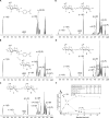
NMR spectroscopic monitoring of the hydrolysis of Galα-pNP by BfGal110Bα-galactosidase. In these one-dimensional 1H NMR spectra of deuterium-exchanged Galα-pNP, only the monosaccharide resonances are shown; downfield aromatic resonances from the nitrophenol are omitted. Spectra were acquired at t = 0(A, prior to enzyme addition), 3 (B), 60 (C), 120 min (D), and 300 min (E) after addition of the enzyme. Structures of predominant species are superimposed on each panel; proton resonance assignments are marked as follows: (S) = signals from substrate, (P) = signals from products, * = probable H-1 from β-furanose form of galactose, HDO = residual monodeuterated water. The reaction time-dependent changes in amplitude of H-1 signals from substrate, α-1 (S), and products, α-1 (P), and β-1 (P), are plotted in F as % of total anomeric (H-1) signal. Inset, table shows progress of reaction for selected time points (min). Tabulated are: approximate α-1 (S) amplitude as a percentage of that at t = 0 (reaction progression), and amplitudes ofα-1 (P) andβ-1 (P) as percentages of total product H-1 signals (productα-1 andβ-1, respectively). Note in F the initial lag in appearance of observable signal forα-1 (P) until after the 25-min time point, whereas that forβ-1 (P) rises rapidly, levels off, and then begins to decline after 140 min, as the amplitudes of the α-1 and β-1 (P) signals approach equilibrium proportions (see Table, inset).
FIGURE 4.
FIGURE 4.
FACS analysis of BfGal110B enzyme-treated porcine and rabbit RBCs. FACS histograms show the relative antigen site density as measured by IB4 (BS-4) lectin on native (solid lines) and enzyme-converted (dashed lines) RBCs from pig (A) or rabbit (B and C). The x-axis represents the fluorescence intensity on a logarithmic scale, whereas the y-axis shows number of RBCs evaluated. The lectin concentration used was 25 μg/ml in A and C, but 5 μg/ml in B. Unstained porcine and rabbit RBCs and human group O RBCs served as additional negative controls and gave similar mean fluorescence intensity values compared with the converted animal RBCs in A and B. In C, higher mean fluorescence intensity was noted for the native rabbit RBCs compared with native porcine RBCs at the same lectin concentration. Post-conversion rabbit RBCs indicate low amounts of remaining Galα1,3Gal epitopes detectable at the higher but not lower lectin concentration shown in C and B, respectively.
FIGURE 5.
FIGURE 5.
TLC analysis of the glycolipids from BfGal110B enzyme-treated rabbit RBCs. Total upper neutral glycolipid fractions from human blood group B RBCs (native B and B-ECO) and rabbit RBCs (native and rabbit ECO) were separated on TLC plates and glycolipids visualized by orcinol staining. Arrows indicate mobilities of major human blood group ABH active glycolipids (B1–B3: Galα1–3(Fucα1–2)(Galβ1–4GlcNAcβ1–3)n = 1–3Galβ1–4Glcβ1-Cer, and H1-H3: Fucα1–2(Galβ1–4GlcNAcβ1–3)n = 1–3Galβ1–4Glcβ1-Cer], rabbit linear B glycolipids (Brab-1–Brab-3: Galα1–3(Galβ1–4GlcNAcβ1–3)n = 1–3Galβ1–4Glcβ1-Cer], Gb4 (globoside: GalNAcβ1–3Galα1–4Galβ1–4Glcβ1-Cer), PG (paragloboside: Galβ1–4GlcNAcβ1–3Galβ1–4Glcβ1-Cer), and nHex (nor-hexosylceramide: Galβ1–4GlcNAcβ1–3Galβ1–4GlcNAcβ1–3Galβ1–4Glcβ1-Cer).

References

    1. Liu, Q. P., Sulzenbacher, G., Yuan, H., Bennett, E. P., Pietz, G., Saunders, K., Spence, J., Nudelman, E., Levery, S. B., White, T., Neveu, J. M., Lane, W. S., Bourne, Y., Olsson, M. L., Henrissat, B., and Clausen, H. (2007) Nat. Biotechnol. 25 454-464 - PubMed
    1. Zhu, A., and Goldstein, J. (1994) Gene (Amst.) 140 227-231 - PubMed
    1. Calcutt, M. J., Hsieh, H. Y., Chapman, L. F., and Smith, D. S. (2002) FEMS Microbiol. Lett. 214 77-80 - PubMed
    1. Davis, M. O., Hata, D. J., Johnson, S. A., Jones, D. E., Harmata, M. A., Evans, M. L., Walker, J. C., and Smith, D. S. (1997) Biochem. Mol. Biol. Int. 42 453-467 - PubMed
    1. Davis, M. O., Hata, D. J., Johnson, S. A., Walker, J. C., and Smith, D. S. (1996) Biochem. Mol. Biol. Int. 39 471-485 - PubMed

Publication types

MeSH terms

LinkOut - more resources