. 2008 May 9;283(19):13194-204.
doi: 10.1074/jbc.M709712200. Epub 2008 Jan 28.

Cytoplasmic terminus of vacuolar type proton pump accessory subunit Ac45 is required for proper interaction with V(0) domain subunits and efficient osteoclastic bone resorption

Affiliations

Cytoplasmic terminus of vacuolar type proton pump accessory subunit Ac45 is required for proper interaction with V(0) domain subunits and efficient osteoclastic bone resorption

Haotian Feng et al. J Biol Chem. .

Abstract

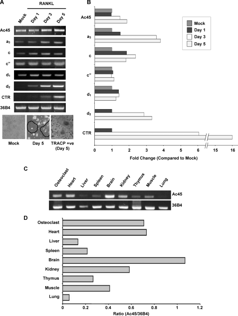
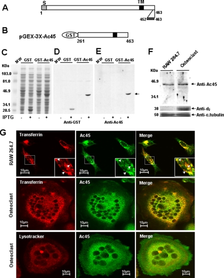
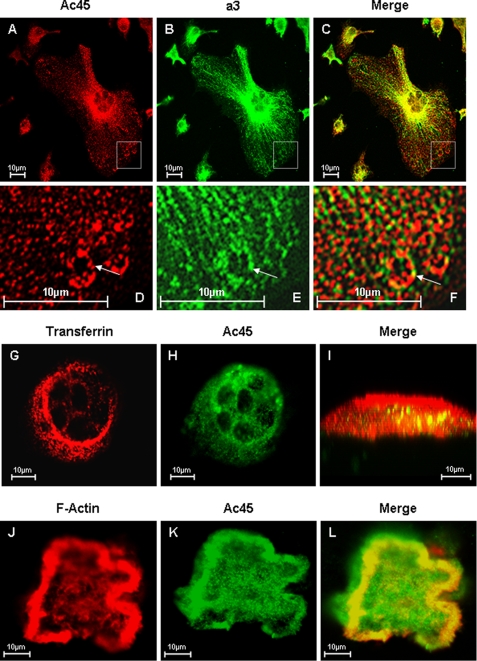
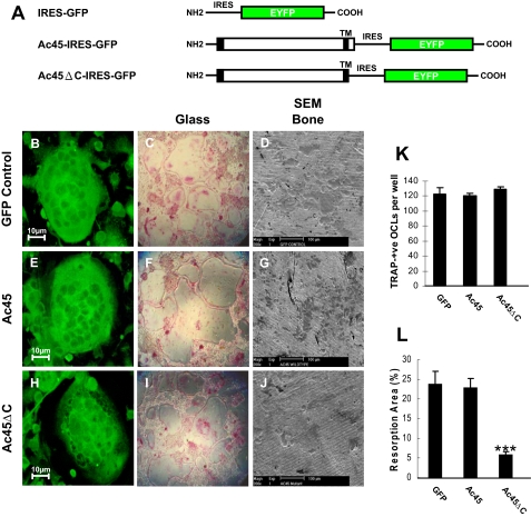
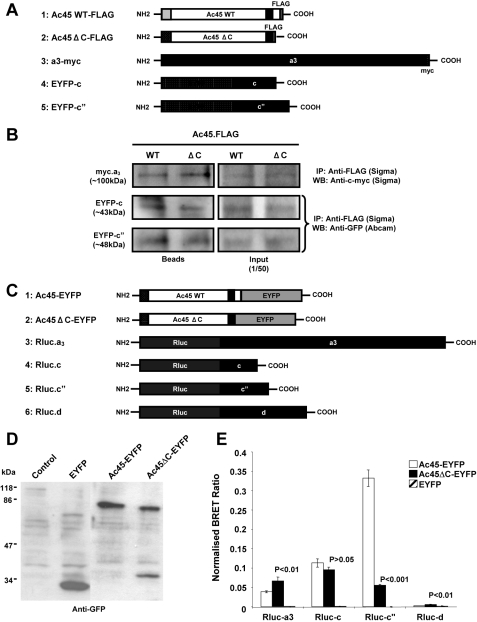
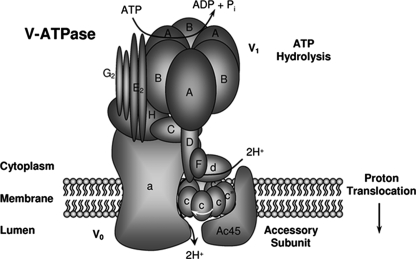
Solubilization of mineralized bone by osteoclasts is largely dependent on the acidification of the extracellular resorption lacuna driven by the vacuolar (H+)-ATPases (V-ATPases) polarized within the ruffled border membranes. V-ATPases consist of two functionally and structurally distinct domains, V(1) and V(0). The peripheral cytoplasmically oriented V(1) domain drives ATP hydrolysis, which necessitates the translocation of protons across the integral membrane bound V(0) domain. Here, we demonstrate that an accessory subunit, Ac45, interacts with the V(0) domain and contributes to the vacuolar type proton pump-mediated function in osteoclasts. Consistent with its role in intracellular acidification, Ac45 was found to be localized to the ruffled border region of polarized resorbing osteoclasts and enriched in pH-dependent endosomal compartments that polarized to the ruffled border region of actively resorbing osteoclasts. Interestingly, truncation of the 26-amino acid residue cytoplasmic tail of Ac45, which encodes an autonomous internalization signal, was found to impair bone resorption in vitro. Furthermore, biochemical analysis revealed that although both wild type Ac45 and mutant were capable of associating with subunits a3, c, c'', and d, deletion of the cytoplasmic tail altered its binding proximity with a3, c'', and d. In all, our data suggest that the cytoplasmic terminus of Ac45 contains elements necessary for its proper interaction with V(0) domain and efficient osteoclastic bone resorption.


Figures

FIGURE 1.
FIGURE 1.
A, different V0 subunits expression profiles during osteoclastogenesis. RAW264.7 cells were treated with RANKL (100 ng/ml) for different time periods (0, 1, 3, and 5 days). RT-PCR analysis was carried out using specific primers to Ac45 and V0 subunits including a3, c, c″, d1, and d2. Calcitonin receptor (CTR) and 36B4 primers were used as control for osteoclastogenesis and house keeping gene, respectively. TRACP staining in a parallel experiment was also included with OCLs highlighted with circles. B, the expression of each subunit relative to 36B4 during osteoclastogenesis was expressed as the fold change over mock. C, RT-PCR analysis of Ac45 (upper panel) and 36B4 (lower panel) in various mouse tissues. D, the relative expression of Ac45 in various tissues relative to 36B4.
FIGURE 2.
FIGURE 2.
Expression and localization of Ac45 in OCLs. A and B, expression of GST-Ac45 and Western analysis with an anti-Ac45 antibody. A, molecular structure of the mouse Ac45. S, signal sequence; TM, transmembrane region. C-terminal amino acid residues 452–463 were used for the generation of anti-Ac45 rabbit polyclonal antibody. B, a cDNA fragment encompassing amino acid residues 261–463 of mouse Ac45 was cloned into the pGEX-3X expression vector. C, Coomassie Blue-stained polyacrylamide gel showing the induction and expression of GST-Ac45261–463 fusion proteins in E. coli. MW, molecular mass. D and E, Western blot analysis of GST fusion proteins using a rabbit anti-GST antibody (D) or a rabbit anti-Ac45 antibody (E). F, Western blot analysis of Ac45 in RAW cell-derived osteoclasts and their precursor cells. A band corresponding to a 45-kDa protein was detected using the Ac45 antibody in both in RAW cell-derived osteoclasts and their precursor cells. Protein expression of V-ATPase subunit d2 was used as positive control, and anti-α-tubulin was used as an internal control. G, confocal microscopy analysis of Ac45 protein localization in osteoclastic precursor RAW264.7 cells and in no-resorbing OCLs. RAW264.7 cells or RAW cell-derived osteoclasts were seeded on glass coverslips. Transferrin Alexa Fluor 546 was added to a final concentration of 50 μg/ml and incubated for 30 min. Lysotracker was added to cell culture at 1 μl/ml and incubated for 30 min. The cells were fixed with 4% paraformaldehyde in PBS and stained with purified anti-Ac45 antibody. Fluorescent-labeled secondary antibody was used, and fluorescent images were recorded using a confocal laser scanning microscope (MRC-1000 Bio-Rad). The cells stained with secondary antibody only were used as the negative control and showed no staining signals.
FIGURE 3.
FIGURE 3.
Confocal microscopy analysis of Ac45 co-localization with a3 in OCLs. AE, osteoclasts cultured on glass coverslips were fixed and permeabilized in 0.1% Triton X-100. The cells were immunostained with primary rabbit Anti-Ac45 and guinea pig anti-a3 for 2 h followed by secondary goat anti-rabbit Alexa Fluor 546 (A) and fluorescein isothiocyanate-conjugated anti-guinea pig (B) for 45 min, respectively. Co-localization was examined following the overlaying of the Ac45 and a3 signals (C). D–F, higher magnification of the boxed area. GI, osteoclasts seeded onto bone slices for 3 days were incubated with Alexa Fluor 546-conjugated transferrin (G) for 30 min followed by fixation and permeabilization. The cells were immunostained with an anti-Ac45 antibody (H) for 2 h and then with secondary goat anti-rabbit Alexa Fluor 488 for 45 min before confocal analysis. Co-localization between Ac45 and transferrin filled endosomal vesicles were observed in z-x or vertical section imaging (I). JL, confocal microscopy analysis of Ac45 protein localization in resorbing osteoclasts. Following 3-day culture on bone slices, osteoclasts were fixed and permeabilized in 0.1% Triton X-100. The cells were again immunostained for with an anti-Ac45 antibody (J) and secondary goat anti-rabbit Alexa Fluor 488. F-actin was stained with rhodamine-conjugated phalloidin (K). Ac45 was observed to extensively co-localize with F-actin surrounding the resorption lacunae (L).
FIGURE 4.
FIGURE 4.
The effect of overexpression of Ac45 or Ac45ΔC on osteoclast formation and osteoclastic bone resorption. A, schematic representation of GFP, Ac45-IRES-GFP, and Ac45ΔC-IRES-GFP constructs. Bone marrow cell derived osteoclasts transduced with GFP (BD), Ac45-IRES-GFP (EG), or Ac45ΔC-IRES-GFP (HJ) retroviruses. Transduced cells were seeded on glass coverslips or 96-well culture plates or bone slices in the presence of RANKL and M-CSF. After 7 days on the glass coverslips or the culture dishes, the cells were fixed and processed for confocal analysis (B, E, and H) or TRACP staining (C, F, and I). In 96-well culture plates, TRACP positive OCLs were counted, and no significant difference was observed between all groups (K). The cells on the bone slices were removed after 14 days of culture, and resorptive lacunae were examined by scanning electron microscopy (D, G, and J). Quantitative analysis shows the percentage of bone slice surface occupied by resorption lacunae (L). ***. p < 0.001.
FIGURE 5.
FIGURE 5.
A, schematic representation of Ac45-FLAG, Ac45ΔC-FLAG, a3-cMyc, EYFP-c, and EYFP-c″ constructs used for immunoprecipitation assays. B, immunoprecipitation analysis showing that Ac45 and Ac45ΔC interact with a3, c, and c″, respectively. C, schematic representation of Ac45-EYFP, Ac45ΔC-EYFP, Rluc-a3, Rluc-c, Rluc-c″, and Rluc-d1 constructs used for BRET assays. D, Western blot analysis of COS-7 cells transfected with EYFP, Ac45-EYFP, or Ac45ΔC. Transfected COS-7 cells were lysed in standard sample buffer, and cell lysates (30 μl) were subjected to analysis by SDS-PAGE and immunoblotting with anti-GFP rabbit polyclonal antibody. E, BRET assays showing the levels of interaction of Ac45 with V0 subunits a, c, c″, and d1. COS-7 cells co-expressing Ac45-EYFP or Ac45ΔC-EYFP and either Rluc tagged a3, c, c″, or d1 subunits were assayed following the addition of coelenterazine and the BRET ratio relative to the Rluc alone expressing cells determined (normalized BRET ratio). Similarly, the normalized BRET ratio was determined for cells co-expressing EYFP with each of the Rluc-tagged subunits. The data represent the means from six independent experiments ± S.E. p values < 0.05 indicate significant differences between Ac45-EYFP and EYFP alone control. p values < 0.05 indicate the significant differences between Ac45 mutant and wild type (WT) Ac45.
FIGURE 6.
FIGURE 6.
Schematic diagram of the predicted localization and close proximity of the Ac45 with other V0 subunits in the V-ATPase complex.

Similar articles

Cited by

References

    1. Teitelbaum, S. L. (2000) Science 289 1504-1508 - PubMed
    1. Blair, H. C., Teitelbaum, S. L., Ghiselli, R., and Gluck, S. (1989) Science 245 855-857 - PubMed
    1. Pavlos, N. J., Xu, J., Riedel, D., Yeoh, J. S. G., Teitelbaum, S. L., Papadimitriou, J. M., Jahn, R., Ross, F. P., and Zheng, M. H. (2005) Mol. Cell. Biol. 25 5253-5269 - PMC - PubMed
    1. Zhao, H., Laitala-Leinonen, T., Parikka, V., and Vaananen, H. K. (2001) J. Biol. Chem. 276 39295-39302 - PubMed
    1. Baron, R., Neff, L., Louvard, D., and Courtoy, P. J. (1985) J. Cell Biol. 101 2210-2222 - PMC - PubMed

Publication types

Substances