. 2008 Apr 15;94(8):3115-25.
doi: 10.1529/biophysj.107.120014. Epub 2008 Jan 28.

Kinesin and dynein-dynactin at intersecting microtubules: motor density affects dynein function

Affiliations

Kinesin and dynein-dynactin at intersecting microtubules: motor density affects dynein function

Jennifer L Ross et al. Biophys J. .

Abstract

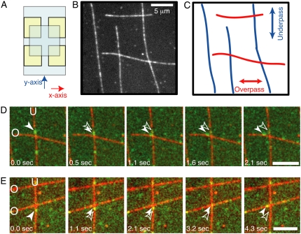
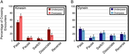
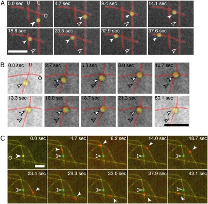
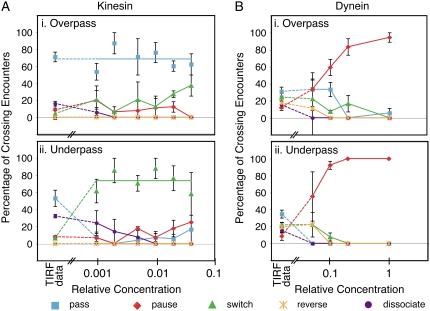
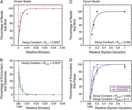
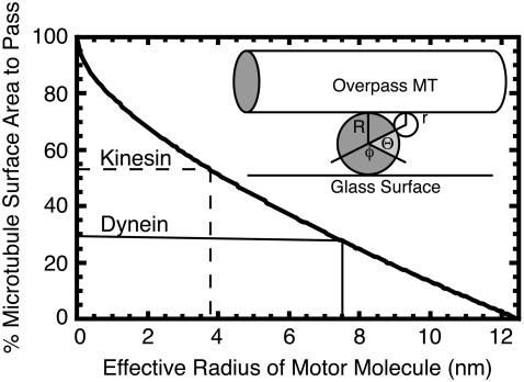
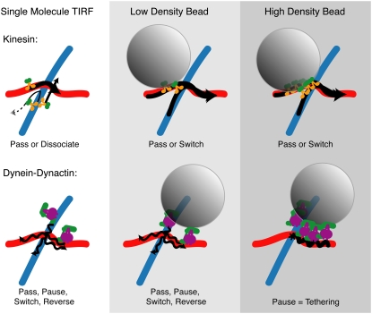
Kinesin and cytoplasmic dynein are microtubule-based motor proteins that actively transport material throughout the cell. Microtubules can intersect at a variety of angles both near the nucleus and at the cell periphery, and the behavior of molecular motors at these intersections has implications for long-range transport efficiency and accuracy. To test motor function at microtubule intersections, crossovers were arranged in vitro using flow to orient successive layers of filaments. Single kinesin and cytoplasmic dynein-dynactin molecules fused with green-fluorescent protein, and artificial bead cargos decorated with multiple motors, were observed while they encountered intersections. Single kinesins tend to cross intersecting microtubules, whereas single dynein-dynactins have a more varied response. For bead cargos, kinesin motion is independent of motor number. Dynein beads with high motor numbers pause, but their actions become more varied as the motor number decreases. These results suggest that regulating the number of active dynein molecules could change a motile cargo into one that is anchored at an intersection, consistent with dynein's proposed transport and tethering functions in the cell.


Figures

FIGURE 1
FIGURE 1
Experimental crossed-flow-path sample chamber and resulting microtubule array. (A) Schematic of crossed-flow-path chamber. The bottom coverglass is 22 mm × 40 mm (pale blue). Four square pieces of double-sided adhesive tape (yellow) are arranged to make two perpendicular 3-mm-wide flow paths. The top coverglass is 18 mm × 18 mm. We denote the flow paths as the x- and y-directions. (B) Example image of rhodamine- and biotin-labeled microtubules bound to the coverglass of a crossed-path flow chamber. Scale bar: 5 μm. (C) Schematic of the same location in B to highlight the crossing microtubule tracks. (D) A single GFP-kinesin, imaged using total internal reflection microscopy, starts walking on the vertical, underpass microtubule (U) and switches to walking on the horizontal, overpass (O) microtubule. (E) A single dynein-dynactin-GFP complex, imaged using total internal reflection fluorescence microscopy, starts walking on the vertical, underpass microtubule (U) and switches to walking on the horizontal, overpass microtubule (O). Green image is GFP fluorescence. Red image is rhodamine fluorescence. Scale bars: 5 μm. See supplementary movies and Fig. S1 for raw images without false coloring.
FIGURE 2
FIGURE 2
Outcomes of GFP-labeled single motor complexes on encountering a microtubule intersection from an overpass or an underpass. (A) Actions of single molecules of kinesin-GFP when encountering an intersection from the underpass (dark red) or overpass (light red) (mean ± SE; n > 60). Kinesin motors mostly pass and dissociate at intersections. (B) Actions of single molecules of dynein-dynactin-GFP when encountering an intersection from the underpass (dark blue) or overpass (light blue) (mean ± SE; n > 30). For dynein-dynactin, all five actions are about equally likely when a complex approaches from an overpass or underpass microtubule.
FIGURE 3
FIGURE 3
Sample time series of motor-decorated bead cargos switching between perpendicular microtubules at an intersection. (A) A bead (false-colored yellow) is coated with multiple kinesin motors and observed to walk along the microtubules (red). The bead is observed to switch from underpass to overpass at the first intersection and then pass by an intersection on an overpass. (B) A bead (false-colored yellow) is coated with multiple dynein-dynactin complexes and observed to walk along the microtubules (red). The bead is observed to pause at an intersection. (C) A bead is coated with multiple kinesin motors and observed to switch tracks and pass at various intersections. In the color overlay, the green image shows the positions of the bead and microtubules at the start of the assay (0.0 s), and the red images are the subsequent ones in the time series. At time points 8.2 and 29.3 s, the microtubules are observed to flex and pivot about the intersection as a result of the kinesin-coated bead interacting strongly with both simultaneously. Each image here consists of four images averaged to highlight microtubule bending. Scale bars: 5 μm. For all images, the white, outlined arrow denotes the starting position, and the filled arrow denotes the position at each time point. The underpass microtubules are marked with a “U”, and the overpass microtubules are marked with an “O” in the first frame. See supplementary movies and Fig. S2 for raw images without false coloring.
FIGURE 4
FIGURE 4
Interactions of kinesin- and dynein-dynactin-decorated beads on encountering intersections plotted versus relative density of motors bound to the bead. (A) Bead cargos coated with kinesin at a variety of decoration densities showed similar statistics when traversing on the (i) overpass or (ii) underpass microtubule. In i the blue line indicates the average percentage of single molecules and beads passing for all kinesin densities. In ii, the green line indicates the average percentage of beads switching for all kinesin densities. (B) Bead cargos coated with dynein-dynactin show a large dependence on motor number when traversing on the overpass (i) or underpass (ii) microtubule. For all plots: passes (blue squares and lines), pauses (red diamonds and lines), switches (green triangles and lines), dissociation (orange stars and lines), and reversals (purple circles and lines); the leftmost symbols denote the single-molecule TIRF data for comparison. Dashed lines are drawn from the TIRF data to the bead data as a guide to the eye.
FIGURE 5
FIGURE 5
Comparison of binding statistics and actions at intersections for kinesin- and dynein-dynactin-coated beads. (A) Percentage of kinesin-coated beads that bound microtubules (red circles) as a function of relative kinesin concentration and fit to the data (red line, χ2 = 22.6). (B) Percentage of kinesin beads that dissociated at the intersection from the underpass (green triangles) as a function of relative kinesin concentration and fit to the data (green line, χ2 = 0.001). (C) Percentage of dynein-dynactin beads that bound microtubules (blue squares) as a function of relative dynein-dynactin and fit to the data (blue line, χ2 = 217). (D) Percentage of dynein-dynactin beads that paused at the intersection from the underpass (open squares) and overpass (solid squares) as a function of relative dynein-dynactin concentration and fits to the data for underpass (dashed line, χ2 = 0.016) and overpass (solid line, χ2 = 0.018). For panels A, C, and D, the fit equation is p = 100 × (1 − exp(−[Motor]/R)), where R is the fit parameter given in the panel. For panel B, the fit equation is: p = pmax × exp(−[Motor]/R), where R is the fit parameter given in the panel.
FIGURE 6
FIGURE 6
Geometric model to estimate the free microtubule circumference for kinesin or dynein to traverse an underpass without hindrance. The percentage of passable surface area is plotted as a function of effective motor radius, r, as explained in the text. When the percentage of passable surface area is 53%, then the effective motor radius is 3.8 nm, as for kinesin (dashed marker lines). When the percentage of passable surface area is 29%, then the effective motor radius is 7.5 nm for dynein (solid marker lines). (Inset) Schematic for the geometric model. The overpass microtubule is represented as a cylinder on top, and the underpass microtubule is seen in cross-section as a circle, both of radius R. The motor is represented as a small circle of radius r. The angle Θ corresponds to the circumference of the underpass microtubule that is accessible to the motor on each side of the microtubule. The angle φ corresponds to the circumference of the underpass microtubule that is permanently blocked by the glass surface.
FIGURE 7
FIGURE 7
Cartoon depiction of the results of single-molecule TIRF and low-density and high-density motor decoration on motility in vitro. Single molecules of kinesin mostly pass and dissociate, whereas single dynein-dynactin complexes can pass, pause, dissociate, switch, and reverse direction (left column). Adding the complexity of a bead cargo with very few motors (middle column) allows kinesin to pass but also to switch frequently. Dynein-dynactin bound to beads at low density allows similar actions, with less passing on an underpass and more pausing, but also some reversing and switching (middle column). Increasing the density of bound kinesin does not significantly change the actions of cargo at intersections, but increasing the density of dynein-dynactin causes all cargo to become tethered at intersections (right column). Wavy lines denote lateral wobbling of the dynein-dynactin bead cargo.

Similar articles

Cited by

References

    1. Vale, R. D. 2003. The molecular motor toolbox for intracellular transport. Cell. 112:467–480. - PubMed
    1. Svoboda, K., C. F. Schmidt, B. J. Schnapp, and S. M. Block. 1993. Direct observation of kinesin stepping by optical trapping interferometry. Nature. 365:721–727. - PubMed
    1. Cai, D., A. D. Hoppe, J. A. Swanson, and K. J. Verhey. 2007. Kinesin-1 structural organization and conformational changes revealed by FRET stoichiometry in live cells. J. Cell Biol. 176:51–63. - PMC - PubMed
    1. Kon, T., M. Nishiura, R. Ohkura, Y. Y. Toyoshima, and K. Sutoh. 2004. Distinct functions of nucleotide-binding/hydrolysis sites in the four AAA modules of cytoplasmic dynein. Biochemistry. 43:11266–11274. - PubMed
    1. Burgess, S. A., M. L. Walker, H. Sakakibara, P. J. Knight, and K. Oiwa. 2003. Dynein structure and power stroke. Nature. 421:715–718. - PubMed

Publication types

MeSH terms

LinkOut - more resources