. 2008 Apr;28(7):2437-45.
doi: 10.1128/MCB.01886-07. Epub 2008 Jan 28.

Trypanosoma brucei RNA editing: coupled cycles of U deletion reveal processive activity of the editing complex

Affiliations

Trypanosoma brucei RNA editing: coupled cycles of U deletion reveal processive activity of the editing complex

Vadim S Alatortsev et al. Mol Cell Biol. 2008 Apr.

Abstract

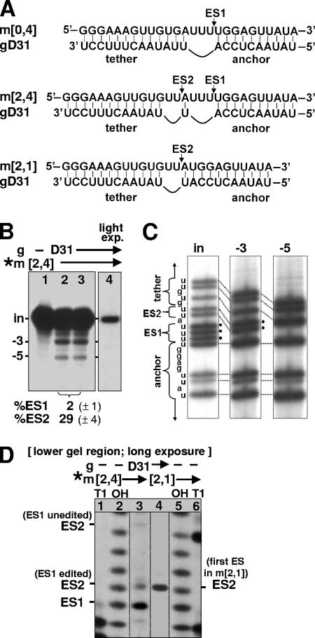
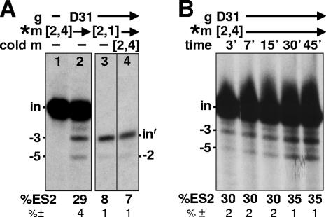
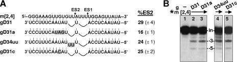
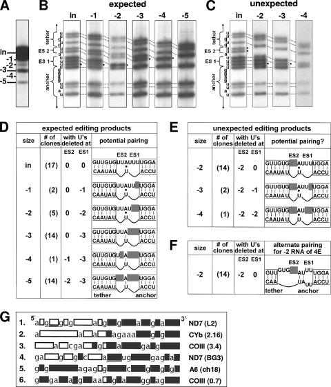
RNA editing in Trypanosoma brucei is posttranscriptional uridylate removal/addition, generally at vast numbers of pre-mRNA sites, but to date, only single editing cycles have been examined in vitro. We here demonstrate achieving sequential cycles of U deletion in vitro, with editing products confirmed by sequence analysis. Notably, the subsequent editing cycle is much more efficient and occurs far more rapidly than single editing cycles; plus, it has different recognition requirements. This indicates that the editing complex acts in a concerted manner and does not dissociate from the RNA substrate between these cycles. Furthermore, the multicycle substrate exhibits editing that is unexpected from a strictly 3'-to-5' progression, reminiscent of the unexpected editing that has been shown to occur frequently in T. brucei mRNAs edited in vivo. This unexpected editing is most likely due to alternate mRNA:guide RNA (gRNA) alignment forming a hyphenated anchor; its having only a 2-bp proximal duplex helps explain the prevalence of unexpected editing in vivo. Such unexpected editing was not previously reported in vitro, presumably because the common use of artificially tight mRNA:gRNA base pairing precludes alternate alignments. The multicycle editing and unexpected editing presented in this paper bring in vitro reactions closer to reproducing the in vivo editing process.


Figures

FIG. 1.
FIG. 1.
Two cycles of U deletion. (A) mRNAs (upper rows) and gRNA (lower rows) used in the in vitro editing reaction. The mRNAs are designated by “m” followed by the number of U's present at ES2 and ES1, respectively, while gRNAs for U deletion are designated by “gD” followed by their nucleotide length, as designated by Cruz-Reyes et al. (12, 14). The mRNA m[0,4] (which has no U's at ES2 and four U's at ES1) is a 72-nt segment from the 3′ region of the natural T. brucei A6 pre-mRNA (12) (it is basically the A6/TAG RNA described by Seiwert and Stuart [44] with its 5′ portion further shortened as described in Seiwert et al. [45]). gD31 is gD30CC (14) with an additional 3′ U, a simplified version of a natural gRNA that from m[0,4] directs deletion of the three unpaired U's (of the four U's present) at ES1 (14) (top). The mRNA m[2,4] contains an additional two U's at ES2 that do not pair with gD31, so this gRNA theoretically could direct deletion of the two U's at ES2 as well as the three U's at ES1 (middle). The mRNA m[2,1] (shown in Fig. 1D and 2) contains two U's at ES2 and one U at ES1, so it is effectively already edited at ES1; it fully base pairs with gD31 at ES1 and thus should exhibit U deletion of only the two unpaired U's at ES2 (bottom). The ES1 and ES2 cleavage sites are indicated by arrows. The lines at the 5′ and 3′ ends of the RNAs represent the following: for pre-mRNAs, 5′-GGAAAGGUUAGGGGGAGGAG AGAAGAAA and ACCUGGCAUC-3′, and for gRNAs, 5′-GGAUAUAC. (B) In vitro editing of radiolabeled (*) m[2,4], using gD31, showing input m[2,4] (in) and the −3 and −5 RNAs (that have lost three and five U's in one or two cycles of U deletion, respectively). Lanes 2 and 3 are duplicate reactions. The values of the measured extent (%) of editing at ES1 and ES2 (see Materials and Methods) are shown at the bottom of the panel along with the standard deviations of those values in parentheses. Lane 4 is a much lighter exposure of lane 3, selected to show the substrate for ES1 editing (the input RNA) as slightly more intense than the substrate for ES2 editing (the −3 band) in lane 3; yet the product from ES2 editing (the −5 band) is readily visible in lane 3, while the product from ES1 editing (the −3 band) is not observed in lane 4. (C) Sequencing (T tracks) of the major class of cDNAs cloned using RNA isolated from gel bands of the indicated sizes, as shown in panel B. The bands in this sequencing represent positions that were U's in the RNA. The anchor and tether regions and the U's to be deleted at ES1 and ES2 are indicated. Dotted lines indicate corresponding residues in the different-length molecules; those residues marked by black circles are deleted in the lane on the right. (D) Cleavage products from double-round U deletion reaction. The lower region of an editing gel, as shown in panel B, using the indicated RNAs (exposure ∼3-fold longer than is optimal for the upper region of the gel). Sizing markers were from each mRNA treated with RNase T1 or with hydroxide (lanes 1, 2, 5, and 6) to generate a G ladder and a nucleotide ladder of that RNA (12, 56). (Since the editing cleavage leaves a 5′ P and the markers a 5′ OH, the bands do not precisely align [references , , and and references therein].) The cleavage at ES2 of m[2,4] that has already been edited at ES1 generates a band that is 1 nt longer than the band from the cleavage at ES1; the cleavage at ES2 of m[2,4] that is unedited at ES1 generates a band that is 4 nt longer than the band from the cleavage at ES1 (A).
FIG. 2.
FIG. 2.
Coupled U deletion cycles. (A) Editing reactions as shown in Fig. 1B, using the indicated RNAs. Lane 3 is a light exposure that shows the substrate for ES2 editing (the m[2,1] input mRNA [in′]) at approximately the same intensity as that of the substrate for ES2 editing in lane 2 (the −3 product from the editing of m[2,4] at ES1). Lane 4 is a normal exposure of a gel lane containing a reaction that used 3% of the input mRNA as radiolabeled (*) m[2,1] and 97% as unlabeled (cold) m[2,4], so its input RNA band is similar in intensity to that shown in lane 3. (B) Kinetics of the editing reaction, with parallel reactions terminated at the indicated times. Note that the substrate and product of ES2 editing in the double-round reaction (the −3 and −5 RNAs, respectively) accumulate in parallel, with an approximately constant percentage of ES2 editing observed at the various times, indicating that ES2 editing occurs rapidly, while ES1 editing occurs more slowly. The measured extent of editing at ES2 (see Materials and Methods) and the standard deviations of those values are shown at the bottom of both panels.
FIG. 3.
FIG. 3.
Effect of gRNA pairing on the coupled editing cycle. (A) m[2,4] paired to gRNA variants that increase (gD31a and gD34uu) or decrease (gD31c) the single-stranded character adjoining ES2. Also shown is the measured extent of editing at ES2 and the standard deviations of those values. (B) Editing reactions as shown in Fig. 1B, using the indicated RNAs.
FIG. 4.
FIG. 4.
Expected and unexpected edited sequences. (A) Gel of double-round U deletion reaction products from m[2,4] and gD31, showing the bands that were cloned as cDNAs. (B and C) Sequencing of cDNAs cloned from editing products of the indicated sizes and comparing to the input size RNA, expanding on the data shown in Fig. 1C to show minor as well as major classes of products. (Fig. 1C showed only the most abundant kind of sequence from the three strongest bands (the input [in], −3, and −5 bands.) The resultant sequences were expected (B) or unexpected (C) from 3′-to-5′ editing of the intended mRNA:gRNA pairing. (D and E) Summary of the sequencing data, both the expected (D) and unexpected (E) editing products. Of the cDNAs with sizes indicated in the first column, the number of clones indicated in parentheses in the second column exhibited U deletion at ES2 and ES1 of the number of residues indicated in the third and fourth columns. (The total number of sequenced clones in each size class does not strictly correspond to the RNA abundance in panel A, which demonstrates that there are fewer −2 mRNAs than −3 mRNAs; however, since many −2 mRNAs represent the unexpected kind of editing, we sequenced more of those clones [hence, more −2-size clones than −3-size clones were sequenced].) The final column shows the sequence of the mRNA (upper rows), with the gray boxes representing deleted U's; the lower rows show the gRNA aligned to maximize pairing with the mRNAs. That pairing would be expected (D) or unexpected (E) to generate the observed editing products. A potential A:U base pair between ES1 and ES2 should form (|) after all three U's at ES1 have been removed (and thus it would help guide the −4 and −5 products in panel D) but should not form otherwise (•); yet, pairing of that mRNA residue should be needed to guide the observed U deletion at ES2 that occurs without that U deletion at ES1 (E). The RNAs extend in 5′ and 3′ directions for an additional 6 nucleotides, as shown in Fig. 1A. Note that although the unexpected editing at ES2 arises at an impressively high frequency, it is considerably less favored than the expected editing at ES1. (F) An alternate mRNA:gRNA alignment that could direct U deletion at ES2 without U deletion at ES1, using a hyphenated anchor, which involves the normal 10-bp anchor duplex plus a 2-bp proximal duplex separated by a 2-nucleotide symmetric bulge. The tether duplex in this mRNA:gRNA alignment contains 9 of the normal 12 bp. (G) Examples of reported in vivo mRNAs that exhibit partial editing. The data in rows 1, 4, and 5 are taken from Koslowsky et al. (27), and the data in rows 2, 3, and 6 are taken from Decker and Sollner-Webb (16); the particular RNA is shown on the right. Letters represent nucleotides that do not become edited; boxes represent U's that should be edited in the mature mRNA and were either edited (gray fill) or not edited (white fill) in that particular sequenced molecule. The editing is mainly U insertions, with U deletions indicated by underlined boxes. Some molecules exhibit the expected 3′-to-5′ progression of editing (rows 1 and 2), while many other molecules reflect unexpected editing, including sites that appear to be edited out of order (rows 3 to 6).

References

    1. Abraham, J. M., J. E. Feagin, and K. Stuart. 1988. Characterization of cytochrome c oxidase III transcripts that are edited only in the 3′ region. Cell 55267-272. - PubMed
    1. Aphasizhev, R., I. Aphasizheva, and L. Simpson. 2003. A tale of two TUTases. Proc. Natl. Acad. Sci. USA 10010617-10622. - PMC - PubMed
    1. Benne, R., J. Van den Burg, J. P. Brakenhoff, P. Sloof, J. H. Van Boom, and M. C. Tromp. 1986. Major transcript of the frameshifted coxII gene from trypanosome mitochondria contains four nucleotides that are not encoded in the DNA. Cell 46819-826. - PubMed
    1. Bhat, G. J., D. J. Koslowsky, J. E. Feagin, B. L. Smiley, and K. Stuart. 1990. An extensively edited mitochondrial transcript in kinetoplastids encodes a protein homologous to ATPase subunit 6. Cell 61885-894. - PubMed
    1. Blum, B., N. Bakalara, and L. Simpson. 1990. A model for RNA editing in kinetoplastid mitochondria: “guide” RNA molecules transcribed from maxicircle DNA provide the edited information. Cell 60189-198. - PubMed

Publication types

LinkOut - more resources