. 2008 Apr;76(4):1379-89.
doi: 10.1128/IAI.00745-07. Epub 2008 Jan 28.

NK cells and gamma interferon coordinate the formation and function of hepatic granulomas in mice infected with the Francisella tularensis live vaccine strain

Affiliations

NK cells and gamma interferon coordinate the formation and function of hepatic granulomas in mice infected with the Francisella tularensis live vaccine strain

Sirosh M Bokhari et al. Infect Immun. 2008 Apr.

Abstract

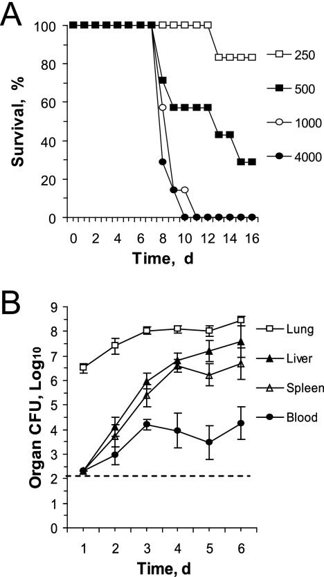
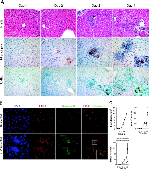
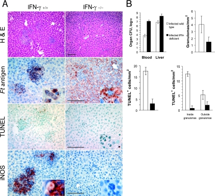
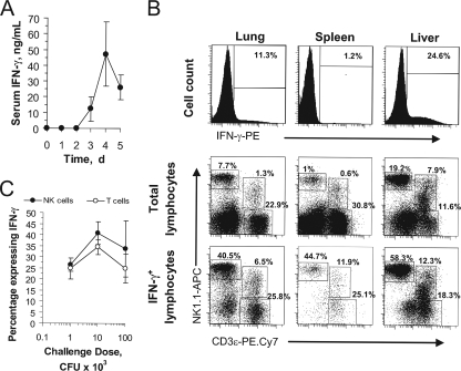
Host innate immune responses to many intracellular pathogens include the formation of inflammatory granulomas that are thought to provide a physical barrier between the microbe and host. Because two common features of infections with the live vaccine strain (LVS) of Francisella tularensis within the mouse liver are the formation of granulomas and the production of gamma interferon (IFN-gamma), we have asked what role IFN-gamma plays in hepatic granuloma formation and function. Francisella antigens were predominantly localized within granulomas of the livers of mice infected with F. tularensis LVS 4 days postinfection. Hepatic granulomas also contained large numbers of dying cells, some of which coexpressed the F4/80 macrophage antigen and activated caspase-3. IFN-gamma-deficient mice did not form normal numbers of hepatic granulomas and showed widely disseminated Francisella antigens within the liver. The incidence of cell death within hepatic granulomas also decreased significantly in the absence of IFN-gamma. Inducible NO synthase (iNOS) expression was restricted to the granulomas of wild-type mice but was not seen for IFN-gamma-deficient mice. Cell death within granulomas was also significantly decreased for iNOS-deficient mice. The predominant IFN-gamma-expressing cells in the liver were NK cells. Depleting NK cells resulted in the expression of bacterial antigens and iNOS outside the granulomas and the appearance of extensive hepatic focal necrosis. These findings indicate that IFN-gamma and hepatic NK cells that are activated during F. tularensis LVS infections regulate hepatic granuloma formation, the spatial containment of infection, the expression of iNOS, and the induction of cell death within the liver.


Figures

FIG. 1.
FIG. 1.
i.n. infection model. (A) To determine the LD50 for mice infected by the i.n. route, C57BL/6J mice (six or seven per group) were challenged with the indicated numbers of viable F. tularensis LVS bacteria. (B) Five mice per group were challenged i.n. with 3.6 × 10³ CFU F. tularensis LVS bacteria, the indicated tissues were collected daily, and total bacterial counts were determined. The horizontal dashed line represents the limit of detection of the assay. d, day.
FIG. 2.
FIG. 2.
Hepatic granuloma formation in F. tularensis LVS-infected mice. (A) Five mice per group were challenged i.n. with F. tularensis LVS and samples of their livers were collected at the indicated times. Fixed tissue samples were stained with hematoxylin and eosin (H&E) to show histopathology, anti-Francisella antigens (Ft antigen) to localize bacterial antigens, or TUNEL reagent to detect cells with double-strand DNA breaks. Note the dying cell with condensed, fragmented chromatin (arrow), the localization of F. tularensis LVS antigens both within the granuloma and occasionally within hepatocytes, and the TUNEL-positive cells concentrated within the granulomas. Bar, 100 μm. (B) Immunofluorescence imaging of a typical hepatic granuloma showing colocalization of activated caspase-3 with the macrophage marker F4/80. The frequency of caspase-3+ cells within granulomas was 27.5 ± 11.2 cells/mm2, while 9.2 ± 5.3 caspase-3+ cells were detected outside the granulomas. (C) The frequencies of granulomas and TUNEL-positive cells increased significantly as a function of time postinfection (P < 0.05) (five mice per group). Granuloma frequency was not scored on day 6 postinfection due to the tendency of granulomas to merge with one another at this time. d, day.
FIG. 3.
FIG. 3.
IFN-γ is required for the formation and function of hepatic granulomas. (A) Wild-type C57BL/6J or coisogenic IFN-γ-deficient mice (four per group) were challenged i.n. with F. tularensis LVS, and their livers were recovered 4 days later. Shown is the effect of IFN-γ deficiency on the localization of Francisella antigens (Ft antigen) and TUNEL-positive cells and the expression of iNOS. H&E, hematoxylin and eosin staining. Bar, 100 μm. (B) Comparison of the bacterial burdens, frequencies of granulomas, and distribution of dying cells within the livers of infected wild-type and IFN-γ-deficient mice. The differences between the wild-type and IFN-γ-deficient mice were significant (P < 0.05) for all measurements, except for the densities of TUNEL+ cells outside the granulomas.
FIG. 4.
FIG. 4.
The expression of iNOS is not required for restricting F. tularensis LVS to hepatic granulomas but does determine the frequency of dying cells in infected livers. (A) Liver samples that were obtained from mice (four mice per group) challenged i.n. with F. tularensis LVS were analyzed for the distribution of Francisella antigens (Ft antigen), TUNEL-positive cells, and iNOS-expressing cells. H&E, hematoxylin and eosin staining. Bars, 100 μm. (B) Tissue bacterial burdens, granuloma frequencies, and frequencies of TUNEL-positive cells in F. tularensis LVS-infected wild-type and iNOS-deficient mice. Compared to wild-type mice, iNOS-deficient mice had significantly low liver bacterial burdens, frequencies of granulomas, and frequencies of TUNEL+ cells both inside and outside the granulomas (P < 0.05).
FIG. 5.
FIG. 5.
The IFN-γ response to i.n. F. tularensis LVS challenge is mediated by pulmonary and hepatic NK cells. (A) Mice (five per group) were challenged i.n. with 3.6 × 10³ CFU F. tularensis LVS bacteria, and circulating IFN-γ levels were measured from serum samples collected on the indicated days postinfection. d, day. (B) Mice were challenged i.n. with F. tularensis LVS, and mononuclear cells were prepared 4 days postchallenge. The cells expressing intracellular IFN-γ were enumerated by flow cytometry (top row). The surface phenotypes of the total mononuclear cells from each organ (middle row) or the IFN-γ-expressing subset of cells (bottom row) were determined by polychromatic flow cytometry. PE, phycoerythrin; APC, allophycocyanin. (C) The percentages of hepatic NK cells or T cells from i.n. challenged mice that expressed intracellular IFN-γ were determined by flow cytometry.
FIG. 6.
FIG. 6.
Depletion of NK cells alters the functions of hepatic granulomas in F. tularensis LVS-infected mice. Wild-type, NK cell-depleted, or T-cell-deficient mice (five per group) were each challenged i.n. with F. tularensis LVS, and samples of their livers were recovered 4 days later. (A) Hepatic mononuclear cells were analyzed by flow cytometry for the expression of intracellular IFN-γ (top row) and the presence of the major subsets of cells (middle row) or the three major subsets of lymphocytes or IFN-γ-producing cells (bottom row). PE, phycoerythrin; APC, allophycocyanin. (B) Granuloma formation and the distribution of CD3ɛ-expressing cells, Francisella antigens (Ft antigen), TUNEL-positive cells, and iNOS in the livers of infected mice. Note the presence of Francisella antigens within hepatocytes of NK cell-depleted mice and the expression of iNOS by Kupffer cells (arrow) and sinusoidal endothelial cells (arrowhead) of these animals. H&E, hematoxylin and eosin staining. (C) Serum IFN-γ concentrations, bacterial counts in the livers, and distribution of TUNEL+ cells in infected mice. Mice depleted of NK cells had significantly more bacteria in their livers, higher totals of TUNEL+ cells, and more TUNEL+ cells outside the granulomas than did wild-type mice (P < 0.05). Mice lacking NK, NKT, and T cells showed a significant decrease in the frequency of granulomas (P < 0.05) and a significant increase in the frequency of TUNEL+ cells in their livers (P < 0.05). (D) Dying cells within the livers of NK cell-depleted mice were found within areas of focal necrosis.

References

    1. Anthony, L. S., E. Ghadirian, F. P. Nestel, and P. A. Kongshavn. 1989. The requirement for gamma interferon in resistance of mice to experimental tularemia. Microb. Pathog. 7421-428. - PubMed
    1. Chen, W., R. KuoLee, H. Shen, and J. W. Conlan. 2004. Susceptibility of immunodeficient mice to aerosol and systemic infection with virulent strains of Francisella tularensis. Microb. Pathog. 36311-318. - PubMed
    1. Cole, L. E., K. L. Elkins, S. M. Michalek, N. Qureshi, L. J. Eaton, P. Rallabhandi, N. Cuesta, and S. N. Vogel. 2006. Immunologic consequences of Francisella tularensis live vaccine strain infection: role of the innate immune response in infection and immunity. J. Immunol. 1766888-6899. - PubMed
    1. Conlan, J. W., W. Chen, H. Shen, A. Webb, and R. KuoLee. 2003. Experimental tularemia in mice challenged by aerosol or intradermally with virulent strains of Francisella tularensis: bacteriologic and histopathologic studies. Microb. Pathog. 34239-248. - PubMed
    1. Conlan, J. W., R. KuoLee, H. Shen, and A. Webb. 2002. Different host defences are required to protect mice from primary systemic vs pulmonary infection with the facultative intracellular bacterial pathogen, Francisella tularensis LVS. Microb. Pathog. 32127-134. - PubMed

Publication types

MeSH terms

LinkOut - more resources