. 2008 Feb 18;205(2):361-72.
doi: 10.1084/jem.20071008. Epub 2008 Jan 28.

Pulmonary arterial remodeling induced by a Th2 immune response

Affiliations

Pulmonary arterial remodeling induced by a Th2 immune response

Eleen Daley et al. J Exp Med. .

Abstract

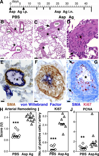
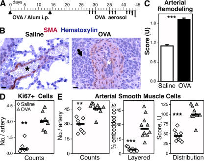
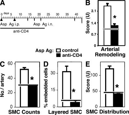
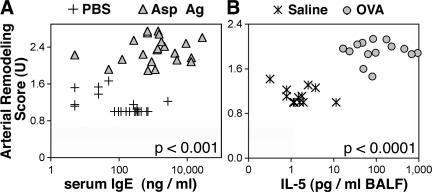
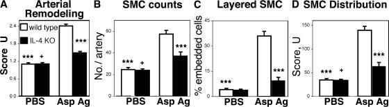
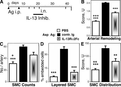
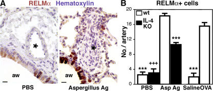
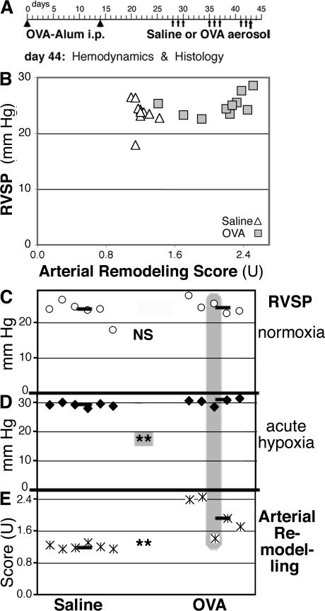
Pulmonary arterial remodeling characterized by increased vascular smooth muscle density is a common lesion seen in pulmonary arterial hypertension (PAH), a deadly condition. Clinical correlation studies have suggested an immune pathogenesis of pulmonary arterial remodeling, but experimental proof has been lacking. We show that immunization and prolonged intermittent challenge via the airways with either of two different soluble antigens induced severe muscularization in small- to medium-sized pulmonary arteries. Depletion of CD4(+) T cells, antigen-specific T helper type 2 (Th2) response, or the pathogenic Th2 cytokine interleukin 13 significantly ameliorated pulmonary arterial muscularization. The severity of pulmonary arterial muscularization was associated with increased numbers of epithelial cells and macrophages that expressed a smooth muscle cell mitogen, resistin-like molecule alpha, but surprisingly, there was no correlation with pulmonary hypertension. Our data are the first to provide experimental proof that the adaptive immune response to a soluble antigen is sufficient to cause severe pulmonary arterial muscularization, and support the clinical observations in pediatric patients and in companion animals that muscularization represents one of several injurious events to the pulmonary artery that may collectively contribute to PAH.


Figures

Figure 1.
Figure 1.
Severe pulmonary arterial remodeling in Asp Ag–exposed mice. (A) Schematic representation of the protocol for priming and challenge (i.p., arrowheads; i.n., arrows) with Asp Ag. (B–G) Lung micrographs from mice exposed to PBS (B and E) or Asp Ag (C, D, F, and G) show H&E staining (B–D) or immunohistochemistry of smooth muscle actin (SMA, brown) and von Willebrand factor (blue; E and F), or SMA (blue) and Ki67 (red; G). Arteries (*), airways (aw), and inflammatory cells (arrows) are indicated. Bars: (B and C) 75 μm; (D and G) 12.5 μm; (E and F) 7.5 μm. Individual data, medians (horizontal lines), and differences between PBS- and Asp Ag–exposed mice (***, P < 0.001; and **, P < 0.01 according to the Wilcoxon U test) are shown for arterial remodeling scores (H) and numbers of Ki67- (I) or PCNA-expressing (J) cells in pulmonary arteries. Data were pooled from two independent experiments.
Figure 2.
Figure 2.
Severe pulmonary arterial remodeling in OVA-exposed mice. (A) Schematic representation of the protocol for priming and challenge (i.p., arrowheads; i.n., arrows) with OVA. (B) Lung micrographs show immunohistochemistry of smooth muscle actin (SMA, red) with hematoxylin (blue) counterstain from saline- or OVA-exposed mice. Arteries (*) and inflammatory cells (arrow) are indicated. Bars: 12.5 μm. (C) Bar graphs show means and SEM of arterial remodeling scores in mice exposed to saline or OVA (***, P < 0.0001 according to the Wilcoxon U test; n = 14–15 mice). Individual data, medians (horizontal lines), and differences between PBS- and OVA-exposed mice (**, P < 0.01; and ***, P < 0.001 according to the Wilcoxon U test) are shown for (D) numbers of Ki67+ cells, and (E) counts, percent layered, and distribution scores of smooth muscle cells in pulmonary arteries. The data were pooled from two independent experiments.
Figure 3.
Figure 3.
Severe pulmonary arterial remodeling is dependent on the presence of CD4+ T cells. (A) Schematic representation of the protocol for CD4 T cell depletion (i.p., arrowheads; i.n., arrows). Bar graphs show means and SEM of (B) scores for arterial remodeling, (C) counts, (D) percent layered, and (E) distribution score of smooth muscle cells (SMC) in pulmonary arteries from mice primed and challenged with Asp Ag and given control or anti-CD4 antibody. Differences between groups (*, P < 0.05 according to the Wilcoxon U test; n = 4 mice) and mean scores determined in PBS-exposed mice (horizontal lines) are indicated.
Figure 4.
Figure 4.
Severe pulmonary arterial remodeling is associated with the development of a Th2 response in Asp Ag– or OVA-exposed mice. Individual data points are shown of arterial remodeling scores plotted against (A) serum IgE levels in mice given PBS or Asp Ag priming and challenge, or (B) BALF IL-5 levels in mice given saline or OVA priming and challenge. The data were analyzed by Spearman's rank correlation test.
Figure 5.
Figure 5.
The IL-4–dependent Th2 response induces severe pulmonary arterial remodeling. Bar graphs show means and SEM of (A) arterial remodeling scores, (B) pulmonary artery smooth muscle cell (SMC) counts, (C) percent layered SMC, and (D) SMC distribution scores from wild-type mice or IL-4KO mice given PBS or Asp Ag priming and challenge. Groups that were significantly different from antigen-primed and -challenged wild-type mice (***, P < 0.001) or from antigen-primed and -challenged IL-4KO mice (+, P < 0.05) are indicated (Bonferroni test; n = 7–10 mice). The data were pooled from two independent experiments.
Figure 6.
Figure 6.
Endogenous IL-13 is critical for pulmonary arterial remodeling. (A) Schematic representation of the protocol for transient IL-13 blockade with IL-13Rα2 mouse Ig constant region (IL-13Rα2-Fc) in mice primed and challenged with Asp Ag. Bar graphs show means and SEM of (B) arterial remodeling scores, (C) pulmonary artery smooth muscle cell (SMC) counts, (D) percent layered SMC, and (E) SMC distribution scores in mice given PBS or Asp Ag priming and challenge and mouse Ig or IL-13Rα2-Fc. Groups that differ from antigen-exposed mice given Ig (**, P < 0.01; ***, P < 0.001) or IL-13Rα2-Fc (+, P < 0.05; ++, P < 0.01; +++, P < 0.001) are indicated (Bonferroni test; n = 9–11 mice). The data are pooled from two independent experiments.
Figure 7.
Figure 7.
Severely remodeled pulmonary arteries are surrounded by RELMα-expressing cells. (A) Lung micrographs show immunohistochemistry of goat anti-RELMα (red) counterstained with hematoxylin (blue) from mice given PBS or Asp Ag priming and challenge. Arteries (*) and airways (aw) are indicated. Bars, 12.5 μm. (B) Bar graphs show means and SEM of counts of RELMα+ cells in wild-type or IL-4KO mice exposed to PBS, Asp Ag, saline, or OVA. Groups different from antigen-primed and challenged wild-type (***) or IL-4KO (+++) mice are indicated (P < 0.001 according to the Bonferroni test; n = 5–10 mice). Data were pooled from two independent experiments for each antigen and mouse strain.
Figure 8.
Figure 8.
Relationship between RVSP and pulmonary arterial remodeling scores. (A) Mice were immunized and exposed to saline or OVA aerosol as shown (i.p., arrowheads; i.n., arrows). (B) Data points show RVSP plotted against arterial remodeling scores in individual mice. The analysis by Spearman's rank correlation test was not significant. Symbols show RVSP in response to (C) normoxia, (D) acute hypoxia (10 min of breathing 10% O2), and (E) corresponding arterial remodeling scores. Data from individual mice are aligned vertically; horizontal bars indicate medians. Statistical analysis was performed with the two-tailed, independent Wilcoxon U test. **, P < 0.01 for the comparison of groups excluding the outlier in the OVA group that did not show severe pulmonary arterial remodeling (outlier data are highlighted in gray; D); **, P < 0.01 for the comparison of groups including all data (E).

References

    1. Heath, D., and J.E. Edwards. 1958. The pathology of hypertensive pulmonary vascular disease; a description of six grades of structural changes in the pulmonary arteries with special reference to congenital cardiac septal defects. Circulation. 18:533–547. - PubMed
    1. Wagenvoort, C.A. 1970. The pathology of primary pulmonary hypertension. J. Pathol. 101:Pi. - PubMed
    1. Reid, L.M. 1979. The pulmonary circulation: remodeling in growth and disease. The 1978 J. Burns Amberson lecture. Am. Rev. Respir. Dis. 119:531–546. - PubMed
    1. Zaiman, A., I. Fijalkowska, P.M. Hassoun, and R.M. Tuder. 2005. One hundred years of research in the pathogenesis of pulmonary hypertension. Am. J. Respir. Cell Mol. Biol. 33:425–431. - PMC - PubMed
    1. Jones, P.L., K.N. Cowan, and M. Rabinovitch. 1997. Tenascin-C, proliferation and subendothelial fibronectin in progressive pulmonary vascular disease. Am. J. Pathol. 150:1349–1360. - PMC - PubMed

Publication types