

Chemical shift assignment of the transmembrane helices of DsbB, a 20-kDa integral membrane enzyme, by 3D magic-angle spinning NMR spectroscopy
- PMID: 18227427
- PMCID: PMC2222720
- DOI: 10.1110/ps.073225008
Chemical shift assignment of the transmembrane helices of DsbB, a 20-kDa integral membrane enzyme, by 3D magic-angle spinning NMR spectroscopy
Abstract
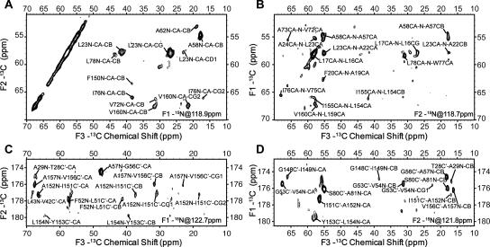
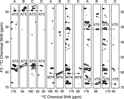
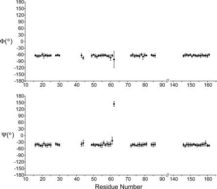
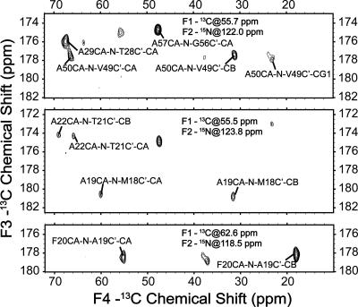
The Escherichia coli inner membrane enzyme DsbB catalyzes disulfide bond formation in periplasmic proteins, by transferring electrons to ubiquinone from DsbA, which in turn directly oxidizes cysteines in substrate proteins. We have previously shown that DsbB can be prepared in a state that gives highly resolved magic-angle spinning (MAS) NMR spectra. Here we report sequential 13C and 15N chemical shift assignments for the majority of the residues in the transmembrane helices, achieved by three-dimensional (3D) correlation experiments on a uniformly 13C, 15N-labeled sample at 750-MHz 1H frequency. We also present a four-dimensional (4D) correlation spectrum, which confirms assignments in some highly congested regions of the 3D spectra. Overall, our results show the potential to assign larger membrane proteins using 3D and 4D correlation experiments and form the basis of further structural and dynamical studies of DsbB by MAS NMR.
Figures
References
-
- Andronesi, O.C., Becker, S., Seidel, K., Heise, H., Young, H.S., Baldus, M. Determination of membrane protein structure and dynamics by magic-angle-spinning solid-state NMR spectroscopy. J. Am. Chem. Soc. 2005;127:12965–12974. - PubMed
-
- Baldus, M., Petkova, A.T., Herzfeld, J., Griffin, R.G. Cross polarization in the tilted frame: Assignment and spectral simplification in heteronuclear spin systems. Mol. Phys. 1998;95:1197–1207.
-
- Bennett, A.E., Rienstra, C.M., Auger, M., Lakshmi, K.V., Griffin, R.G. Heteronuclear decoupling in rotating solids. J. Chem. Phys. 1995;103:6951–6958.
-
- Byrne, B., Iwata, S. Membrane protein complexes. Curr. Opin. Struct. Biol. 2002;12:239–243. - PubMed
-
- Cornilescu, G., Delaglio, F., Bax, A. Protein backbone angle restraints from searching a database for chemical shift and sequence homology. J. Biomol. NMR. 1999;13:289–302. - PubMed
Publication types
MeSH terms
Substances
Grants and funding
LinkOut - more resources
Full Text Sources
