

Editorial
doi: 10.1007/s00417-007-0743-x.
Epub 2008 Jan 29.
Vitreoschisis
- PMID: 18228032
- PMCID: PMC2258312
- DOI: 10.1007/s00417-007-0743-x
Item in Clipboard
Editorial
Vitreoschisis
Graefes Arch Clin Exp Ophthalmol.
2008 Mar.
No abstract available
Figures
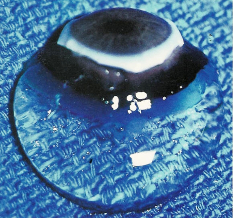
Human vitreous. The sclera, choroid, and retina were dissected off the vitreous in this autopsy specimen obtained form a 9-month-old girl. The vitreous remains attached to the anterior segment. Although the specimen rests upon a surgical towel in room air, the exquisite gel structure is maintained, owing to the young age of the donor. (Specimen courtesy of the New England Eye Bank)
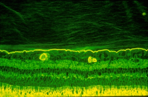
Vitreo–retinal interface. Imunohistochemical studies of the monkey vitreo–retinal interface employed fixation in 4% paraformaldehyde and staining with fluorescein-conjugated ABA lectin. The retina is at the bottom and the vitreous is at the top of this image. The intensely-stained, horizontal linear structure is the internal limiting lamina (ILL) of the retina. Above the ILL is the posterior vitreous cortex. The lamellar structure is clearly evident. Effective magnification ∼400x. (Courtesy of Greg Hageman, Ph.D.)
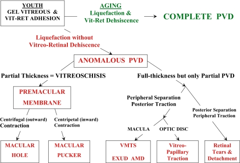
Anomalous PVD. This schematic diagram demonstrates the various possible manifestations of anomalous PVD [10]. When gel liquefaction and weakening of vitreo–retinal adhesion occur concurrently, the vitreous separates away from the retina without sequelae (top of diagram). If the separation of vitreous from retina is full-thickness but incomplete, there can be different forms of partial PVD (right side of diagram). Posterior separation with persistent peripheral vitreo–retinal attachment can induce retinal breaks and detachments. Peripheral vitreo–retinal separation with persistent full-thickness attachment of vitreous to the retina posteriorly can induce traction upon the macula, known as the vitreo–macular traction syndrome (VMTS). This phenomenon appears to be highly associated with exudative AMD [11]. Persistent attachment to the optic disc can induce vitreo-papillopathies and also contribute to neovascularization and vitreous hemorrhage in ischemic retinopathies. If during PVD the posterior vitreous cortex splits (vitreoschisis), there can be differences depending upon the level of the split. Vitreoschsis anterior to the level of the hyalocytes leaves a relatively thick, cellular membrane attached to the macula. Inward (centripetal) contraction of this membrane induces macular pucker. If the split occurs at a level posterior to the hyalocytes, the remaining premacular membrane is relatively thin and hypocellular. Outward (centrifugal) tangential traction can induce a macular hole
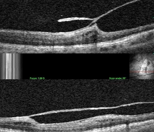
Clinical vitreoschisis. Combined OCT/SLO imaging detected a split in the posterior vitreous cortex. In these two cases, the outer layer of the split posterior vitreous cortex remains adherent to the retina. The point where the two layers re-join into one full-thickness layer is often the site of significant traction upon the retina. Studies have shown that about half of patients with macular hole and macular pucker have vitreoschisis
References
-
- Sebag J (1989) The vitreous—structure, function, and pathobiology. Springer-Verlag, New York
-
- Sebag J (1998) Macromolecular structure of vitreous. Prog Polym Sci 23:415–446
-
- Sebag J (1991) Age-related differences in the human vitreo-retinal interface. Arch Ophthalmol 109:966–971 - PubMed
Publication types
MeSH terms
LinkOut - more resources
Full Text Sources
Medical
