

Fluorescent labeled anti-EGFR antibody for identification of regional and distant metastasis in a preclinical xenograft model
- PMID: 18228526
- PMCID: PMC4402971
- DOI: 10.1002/hed.20782
Fluorescent labeled anti-EGFR antibody for identification of regional and distant metastasis in a preclinical xenograft model
Abstract
Background: Detection of regional and distant metastatic disease has significant implications for patient management. Fluorescent imaging may be a useful technique for metastasis detection and removal.
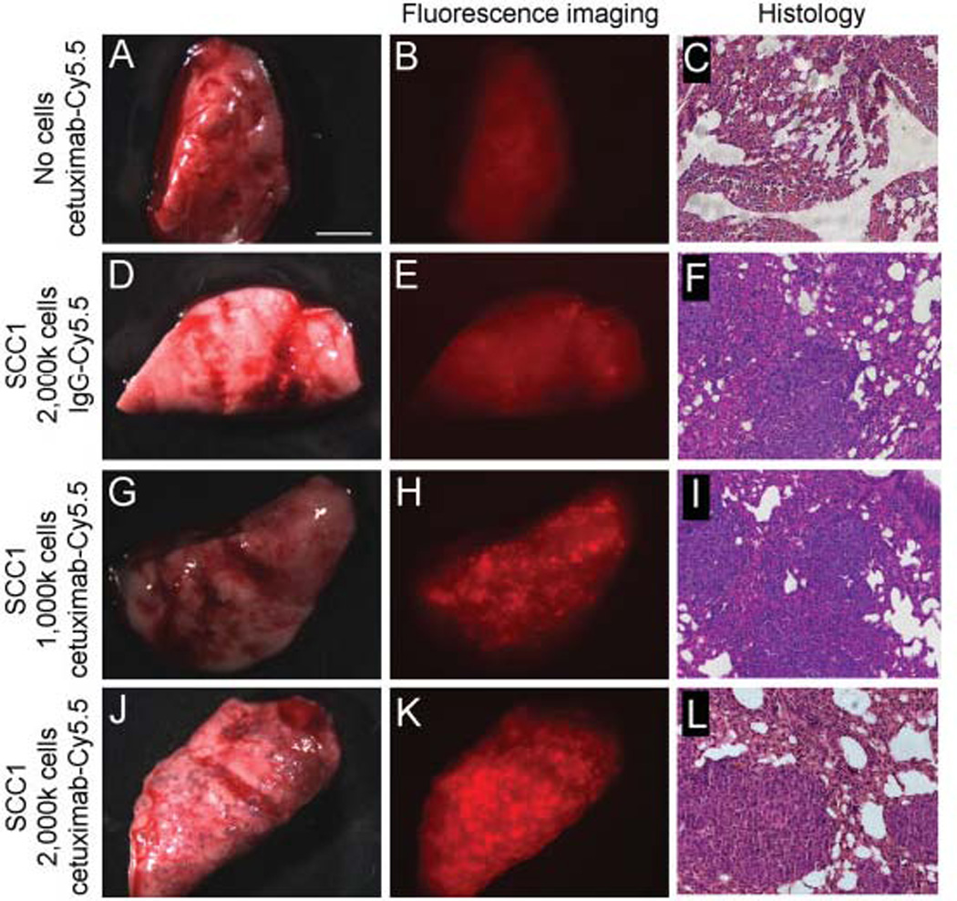
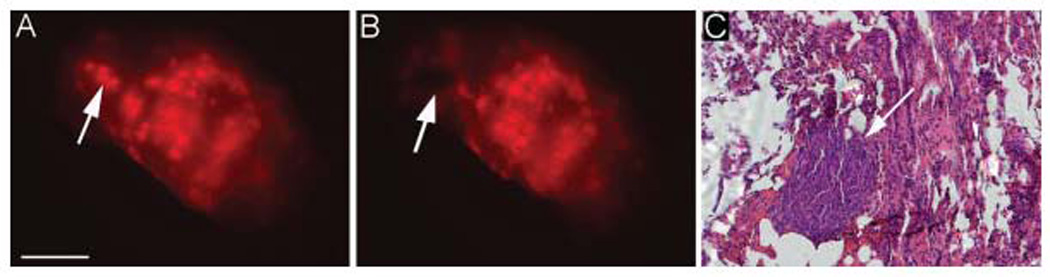
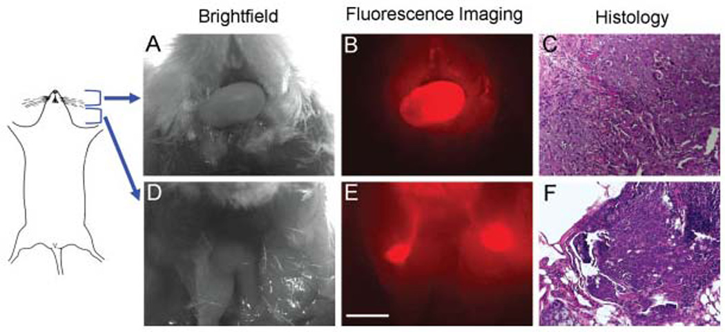
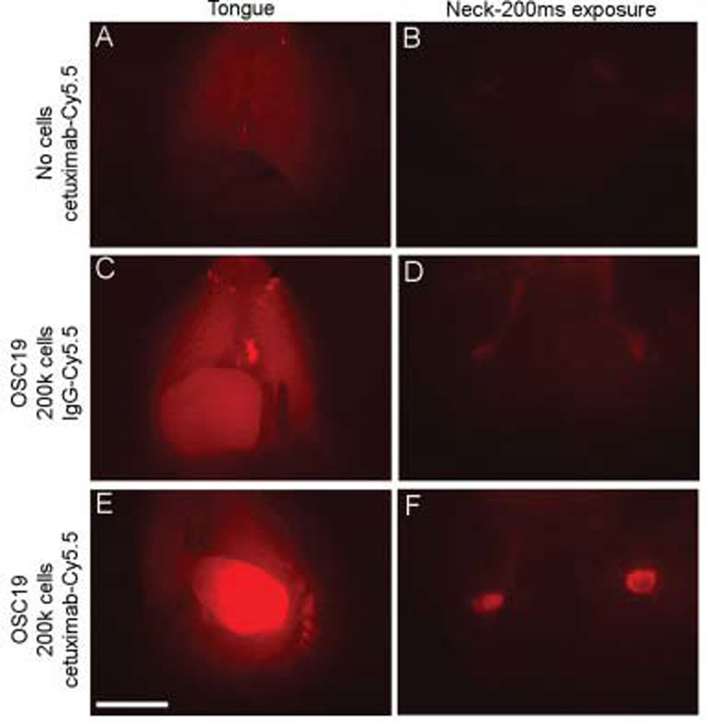
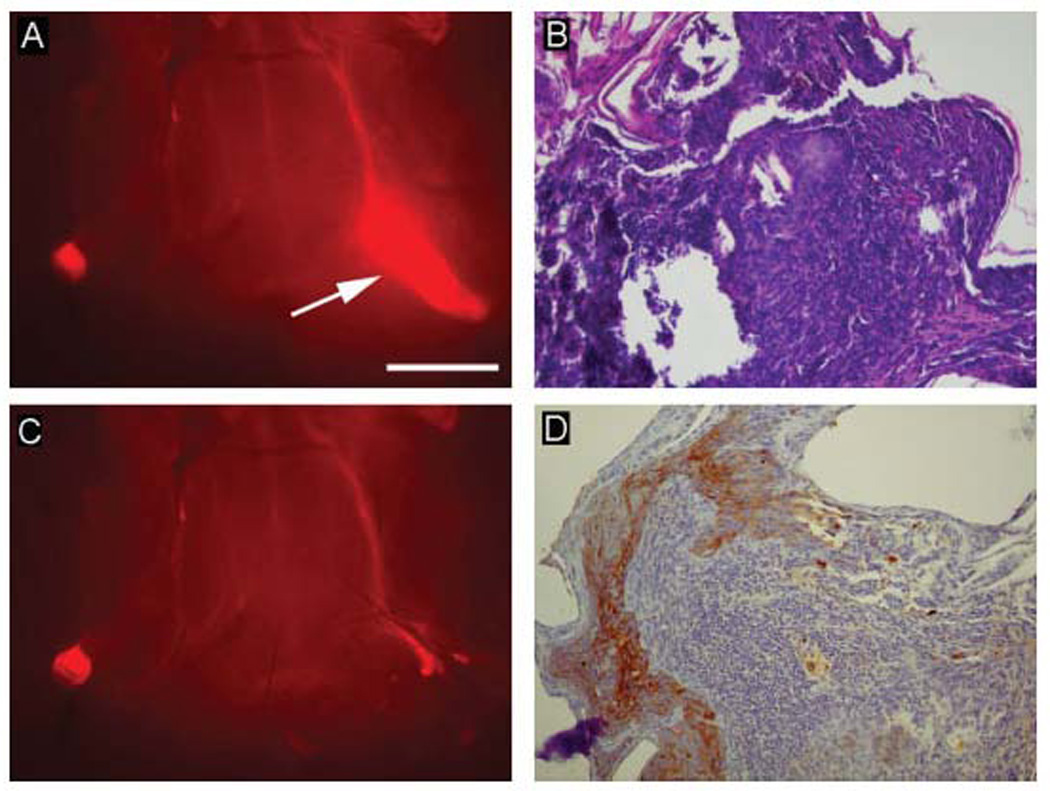
Methods: Anti-epidermal growth factor receptor antibody (cetuximab) and isotype-matched control antibody (immunoglobulin G [IgG]) were labeled with a near-infrared fluorophore (Cy5.5), then systemically administered to mice with tumors resulting from either intraoral or intravenous injections of head and neck squamous cell carcinoma. Mice were sacrificed before undergoing fluorescent stereomicroscopy to assess pulmonary or cervical lymph node metastasis. Fluorescent areas were serially excised until wound bed demonstrated negative fluorescence.
Results: Mice bearing pulmonary metastases displayed diffuse background after IgG-Cy5.5 injection, but demonstrated a speckled fluorescent pattern across lung surface following cetuximab-Cy5.5 injection. Mice bearing cervical metastases demonstrated clear fluorescence of primary tongue tumor and bilateral cervical nodes. Fluorescence correlated with histopathology.
Conclusion: These data suggest that cetuximab-Cy5.5 may have clinical utility in the detection and guided the removal of regional and distant micrometastasis.
Figures
References
-
- Som PM. Detection of metastasis in cervical lymph nodes: CT and MR criteria and differential diagnosis. Am J Roentgenol. 1992;158:961–969. - PubMed
-
- Lardinois D, Weder W, Hany TF, et al. Staging of nonsmall-cell lung cancer with integrated positron-emission tomography and computed tomography. N Engl J Med. 2003;348:2500–2507. - PubMed
-
- Veiseh M, Gabikian P, Bahrami SB, et al. Tumor paint: a chlorotoxin:Cy5.5 bioconjugate for intraoperative visualization of cancer foci. Cancer Res. 2007;67:6882–6888. - PubMed
-
- Koyama Y, Hama Y, Urano Y, Nguyen DM, Choyke PL, Kobayashi H. Spectral fluorescence molecular imaging of lung metastases targeting HER2/neu. Clin Cancer Res. 2007;13:2936–2945. - PubMed
-
- Pomerantz RG, Grandis JR. The role of epidermal growth factor receptor in head and neck squamous cell carcinoma. Curr Oncol Rep. 2003;5:140–146. - PubMed
Publication types
MeSH terms
Substances
Grants and funding
LinkOut - more resources
Full Text Sources
Other Literature Sources
Medical
Research Materials
Miscellaneous
