New insights on COPD imaging via CT and MRI
- PMID: 18229568
- PMCID: PMC2695207
New insights on COPD imaging via CT and MRI
Abstract
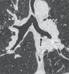
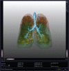
Multidetector-row computed tomography (MDCT) can be used to quantify morphological features and investigate structure/function relationship in COPD. This approach allows a phenotypical definition of COPD patients, and might improve our understanding of disease pathogenesis and suggest new therapeutical options. In recent years, magnetic resonance imaging (MRI) has also become potentially suitable for the assessment of ventilation, perfusion and respiratory mechanics. This review focuses on the established clinical applications of CT, and novel CT and MRI techniques, which may prove valuable in evaluating the structural and functional damage in COPD.
Figures


Similar articles
-
Time-series hyperpolarized xenon-129 MRI of lobar lung ventilation of COPD in comparison to V/Q-SPECT/CT and CT.Eur Radiol. 2019 Aug;29(8):4058-4067. doi: 10.1007/s00330-018-5888-y. Epub 2018 Dec 14. Eur Radiol. 2019. PMID: 30552482 Free PMC article.
-
Oxygen-enhanced magnetic resonance imaging versus computed tomography: multicenter study for clinical stage classification of smoking-related chronic obstructive pulmonary disease.Am J Respir Crit Care Med. 2008 May 15;177(10):1095-102. doi: 10.1164/rccm.200709-1322OC. Epub 2008 Feb 14. Am J Respir Crit Care Med. 2008. PMID: 18276941 Clinical Trial.
-
Imaging phenotypes of chronic obstructive pulmonary disease.J Magn Reson Imaging. 2010 Dec;32(6):1340-52. doi: 10.1002/jmri.22376. J Magn Reson Imaging. 2010. PMID: 21105139 Review.
-
This is what COPD looks like.Respirology. 2016 Feb;21(2):224-36. doi: 10.1111/resp.12611. Epub 2015 Aug 26. Respirology. 2016. PMID: 26333307 Review.
-
Using pulmonary imaging to move chronic obstructive pulmonary disease beyond FEV1.Am J Respir Crit Care Med. 2014 Jul 15;190(2):135-44. doi: 10.1164/rccm.201402-0256PP. Am J Respir Crit Care Med. 2014. PMID: 24873985 Review.
Cited by
-
Probing Changes in Lung Physiology in COPD Using CT, Perfusion MRI, and Hyperpolarized Xenon-129 MRI.Acad Radiol. 2019 Mar;26(3):326-334. doi: 10.1016/j.acra.2018.05.025. Epub 2018 Aug 5. Acad Radiol. 2019. PMID: 30087065 Free PMC article.
-
Imaging in alpha-1 antitrypsin deficiency: a window into the disease.Ther Adv Chronic Dis. 2021 Jul 29;12_suppl:20406223211024523. doi: 10.1177/20406223211024523. eCollection 2021. Ther Adv Chronic Dis. 2021. PMID: 34408834 Free PMC article. Review.
-
Pulmonary Shunt in Critical Care: A Comprehensive Review of Pathophysiology, Diagnosis, and Management Strategies.Cureus. 2024 Sep 3;16(9):e68505. doi: 10.7759/cureus.68505. eCollection 2024 Sep. Cureus. 2024. PMID: 39364515 Free PMC article. Review.
-
Guidelines for diagnosis and management of chronic obstructive pulmonary disease: Joint ICS/NCCP (I) recommendations.Lung India. 2013 Jul;30(3):228-67. doi: 10.4103/0970-2113.116248. Lung India. 2013. PMID: 24049265 Free PMC article.
-
Time-series hyperpolarized xenon-129 MRI of lobar lung ventilation of COPD in comparison to V/Q-SPECT/CT and CT.Eur Radiol. 2019 Aug;29(8):4058-4067. doi: 10.1007/s00330-018-5888-y. Epub 2018 Dec 14. Eur Radiol. 2019. PMID: 30552482 Free PMC article.
References
-
- Albert MS, Cates GD, Driehuys B, et al. Biological magnetic resonance imaging using laserpolarized 129Xe. Nature. 1994;370:199–201. - PubMed
-
- Amundsen T, Torheim G, Waage A, et al. Perfusion magnetic resonance imaging of the lung: characterization of pneumonia and chronic obstructive pulmonary disease. A feasibility study. J Magn Reson Imaging. 2000;12:224–31. - PubMed
-
- Aziz ZA, Wells AU, Desai SR, et al. Functional impairment in emphysema: contribution of airway abnormalities and distribution of parenchymal disease. AJR Am J Roentgenol. 2005;185:1509–15. - PubMed
-
- Bankier AA, De Maertelar V, Keyzer C, et al. Pulmonary emphysema: subjective visual grading versus objective quantification with macroscopic morphometry and thin-section CT densitometry. Radiology. 1999;211:851–8. - PubMed
-
- Bayat S, Le DG, Porra L, et al. Quantitative functional lung imaging with synchrotron radiation using inhaled xenon as contrast agent. Phys Med Biol. 2001;46:3287–99. - PubMed
Publication types
MeSH terms
LinkOut - more resources
Full Text Sources
Other Literature Sources
Medical

