

doi: 10.1016/j.bbrc.2008.01.093.
Epub 2008 Jan 28.
Activation of p53-dependent responses in tumor cells treated with a PARC-interacting peptide
Affiliations
- PMID: 18230339
- PMCID: PMC7398582
- DOI: 10.1016/j.bbrc.2008.01.093
Item in Clipboard
Activation of p53-dependent responses in tumor cells treated with a PARC-interacting peptide
Biochem Biophys Res Commun.
.
Abstract
We tested the activity of a p53 carboxy-terminal peptide containing the PARC-interacting region in cancer cells with wild type cytoplasmic p53. Peptide delivery was achieved by fusing it to the TAT transduction domain (TAT-p53-C-ter peptide). In a two-hybrid assay, the tetramerization domain (TD) of p53 was necessary and sufficient to bind PARC. The TAT-p53-C-ter peptide disrupted the PARC-p53 complex. Peptide treatment caused p53 nuclear relocation, p53-dependent changes in gene expression and enhancement of etoposide-induced apoptosis. These studies suggest that PARC-interacting peptides are promising candidates for the enhancement of p53-dependent apoptosis in tumors with wt cytoplasmic p53.
Figures
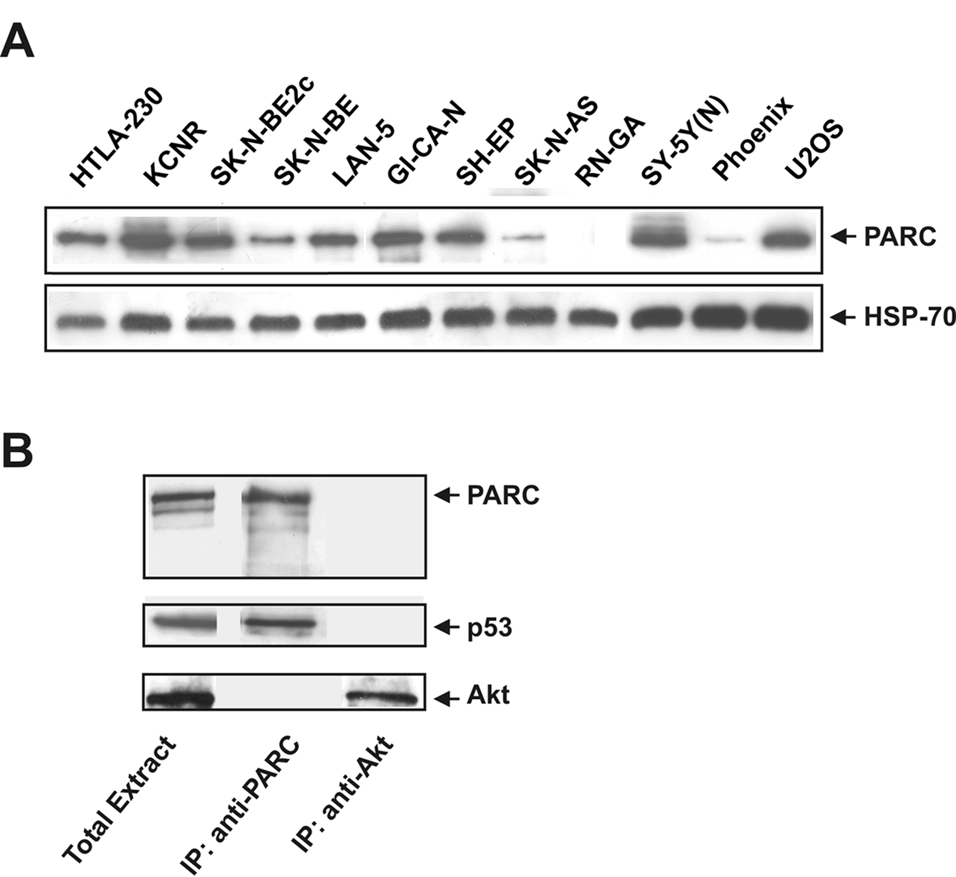
A: endogenous PARC expression in U2OS, Phoenix and 10 human NB cell lines. HSP-70 was used for normalization. B: p53 coimmunoprecipitates with PARC. PARC was immunoprecipitated from LAN-5 cell lysate using an anti-PARC antibody. Control immunoprecipitation was performed using an anti-Akt antibody. Western blots were performed on immunoprecipitates to detect PARC, p53 and Akt.
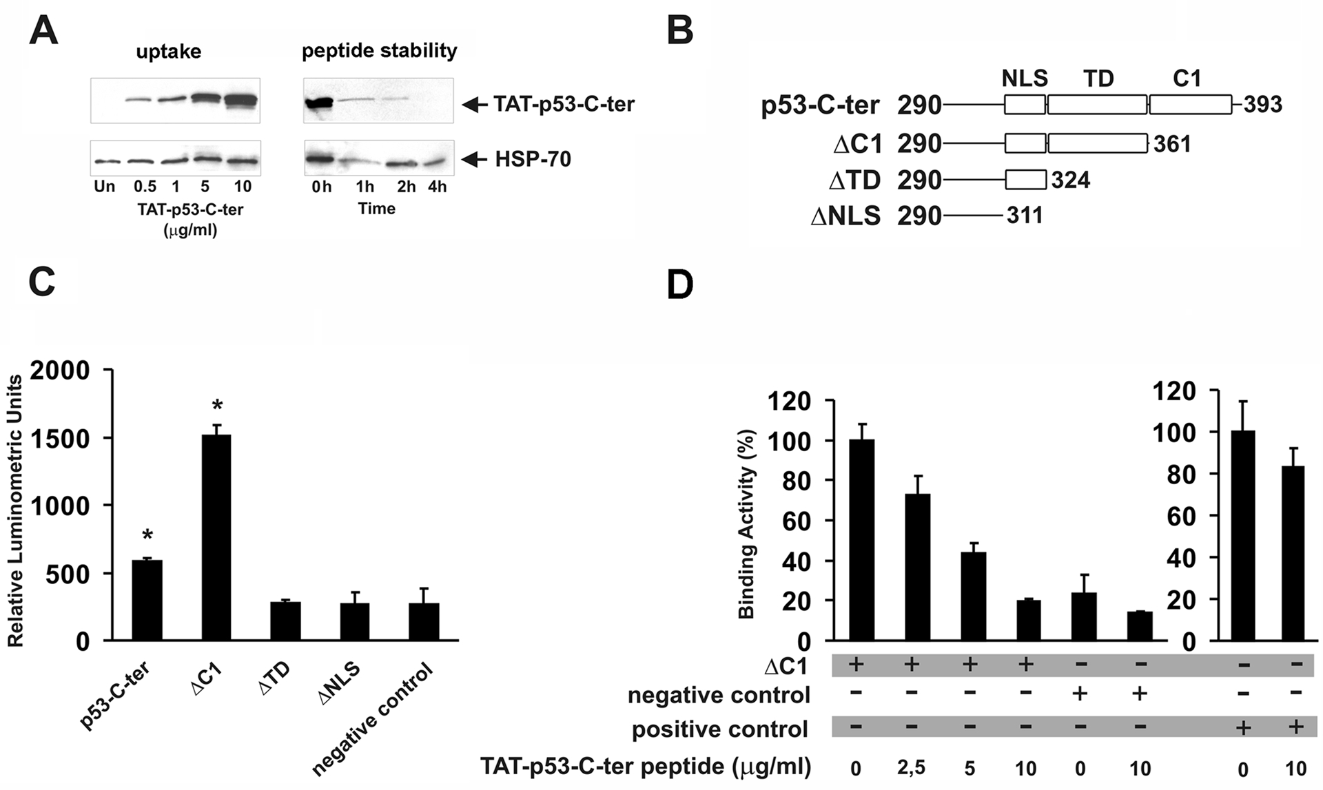
A: dose-dependent uptake of the TAT-p53-C-ter peptide. Cells were treated for 1 hour with the indicated peptide amounts; intracellular TAT-p53-C-ter peptide stability in cells treated with medium containing 10 μg/ml peptide for 1 hour (0h) and in cells cultured for the indicated times in peptide-free medium. In A, peptides were detected with an anti-HA antibody. HSP-70 content was used for normalization. Blots are representative of two different experiments with similar results. B: representation of p53 peptides cloned in the pFN11A vector. NLS= Nuclear Localization Signal; TD= Tetramerization Domain; C1= C1 peptide. Numbers refer to the aminoacids of human p53 protein cloned in each plasmid. C: PARC/p53 binding assay. Phoenix cells were transfected with the reporter vector pGL4.31, pFN10A-CPH-PARC and the pFN11A plasmids containing the p53 peptides described in B. Thirty-six hours after transfection, luciferase activity was determined. Negative control was carried out by transfecting Phoenix cells with pGL4.31 and pACT and pBIND non-interacting vectors. D: Phoenix cells were transfected with pGL4.31, pFN10A-CPH-PARC and pFN11A-ΔC1 plasmids and treated for 6 hours with the indicated amounts of TAT-p53-C-ter peptide (administration every 2 hours) before luminometric detection. Binding activity of untreated cells was taken as 100. Negative control experiments were designed by transfecting Phoenix cells with pGL4.31 and pACT and pBIND non-interacting vectors. As positive control (right histogram), Phoenix cells were transfected with pGL4.31 and pACT-MyoD and pBIND-Id interacting vectors. Binding activity of positive control left untreated was taken as 100. Samples in C and D were run in triplicate. Values ± SD are reported. Asterisks indicate statistically significant differences (p<0.05) compared to negative control. Experiments were repeated twice with similar results.

A: 32D-BCR/ABL myeloid precursor cells stably transfected with reporter vector PG13 or MG15 (luciferase gene driven by a promoter with wt or mutated p53 binding sites respectively) were treated every 2 hours for 6 hours with 5 μg/ml of TAT-p53-C-ter peptide. As positive control, cells were treated for 16 hours with 0.2 μg/ml doxorubicin. B: human NB LAN-5 cells were co-transfected with reporter vectors PG13 or MG15 and expression vector pcDNA3-p53-C-ter-HA. Luciferase activity was determined 36 hours after transfection. As positive control, cells transfected with PG13 and empty vector pcDNA3 were treated for 16 hours with 2 μg/ml doxorubicin. To evaluate additive effects of the p53-C-ter peptide and doxorubicin, cells transfected with PG13 and pcDNA3-p53-C-ter-HA were treated for 16 hours with doxorubicin at the same concentration indicated above. Samples were run in triplicate. Values ± SD are reported. C: Cellular localization of endogenous p53 and TAT-p53-C-ter peptide. U2OS cells, untreated (a, b, c, d), or treated for 1 hour with 10 μg/ml TAT-p53-C-ter peptide (e, f, g and h), or for 1 hour with the peptide followed by 1 hour in peptide-free medium (i, j, k, l), were fixed and processed for immunodetection of endogenous p53 (b, f and j) or the TAT-p53-C-ter peptide (c, g and k). Nuclei were stained with DAPI (a, e and i). Merged fluorescences are shown in d, h and l. Inset in d represents a negative control carried out by treating the cells only with secondary antibodies.
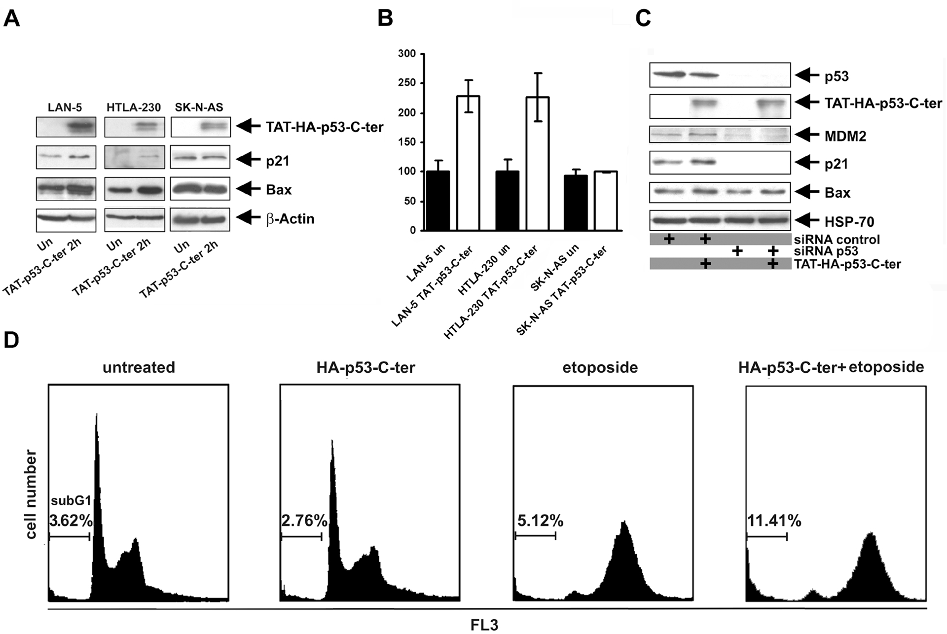
p53 transcription targets are up-regulated by the TAT-p53-C-ter peptide. A: indicated cell lines were treated for 2 hours with 10 μg/ml TAT-p53-C-ter peptide added every hour to the medium. Immunodetection was carried out with the anti-HA (to detect TAT-p53-C-ter peptide), anti-p21, anti-Bax and anti-β-actin antibodies. B: total RNA was extracted from cells treated for 2 hours with 10 μg/ml TAT-p53-C-ter peptide. Quantitative real-time PCR was performed to detect MDM2 expression. Values are normalized for β-actin transcripts of each sample. Reactions were run in triplicate, values ± SD are reported. C: Imunodetection of p53 targets in U2OS cells transfected with p53-siRNA or control-siRNA, untreated or treated with TAT-p53-C-ter peptide (see text for details). D: Flow cytometric analysis of U2OS cells transfected with pcDNA3-p53-C-ter-HA, or control pcDNA3 and pCMVEGFP (4:1) vectors. 24 hours after transfection, cells were treated for 36 hours with 1 μM etoposide or left untreated before fixation and processing for cytometric evaluation. DNA content analyses were performed by gating 20 × 104 green fluorescent cells. Experiments were repeated twice with similar results.
References
-
- Soussi T, Kato S, Levy PP, Ishioka C, Reassessment of the TP53 mutation database in human disease by data mining with a library of TP53 missense mutations Hum. Mutat 25 (2005) 6–17. - PubMed
-
- Nikolaev AY, Li M, Puskas N, Qin J, Gu W, Parc: a cytoplasmic anchor for p53 Cell 112 (2003) 29–40. - PubMed
-
- Kasper JS, Arai T, DeCaprio JA, A novel p53-binding domain in CUL7 Biochem. Biophys. Res. Commun 348 (2006) 132–138. - PubMed
-
- Vassilev LT, Vu BT, Graves B, Carvajal D, Podlaski F, Filipovic Z, Kong N, Kammlott U, Lukacs C, Klein C, Fotouhi N, Liu EA, In vivo activation of the p53 pathway by small-molecule antagonists of MDM2 Science 303 (2004) 844–848. - PubMed
Publication types
MeSH terms
Substances
Grants and funding
LinkOut - more resources
Full Text Sources
Molecular Biology Databases
Research Materials
Miscellaneous
