

Visualization of microtubule growth in living platelets reveals a dynamic marginal band with multiple microtubules
- PMID: 18230754
- PMCID: PMC2343595
- DOI: 10.1182/blood-2007-10-118844
Visualization of microtubule growth in living platelets reveals a dynamic marginal band with multiple microtubules
Abstract
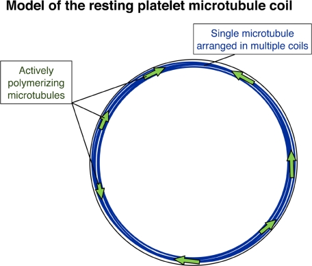
The marginal band of microtubules maintains the discoid shape of resting blood platelets. Although studies of platelet microtubule coil structure conclude that it is composed of a single microtubule, no investigations of its dynamics exist. In contrast to previous studies, permeabilized platelets incubated with GTP-rhodamine-tubulin revealed tubulin incorporation at 7.9 (+/- 1.9) points throughout the coil, and anti-EB1 antibodies stained 8.7 (+/- 2.0) sites, indicative of multiple free microtubules. To pursue this result, we expressed the microtubule plus-end marker EB3-GFP in megakaryocytes and examined its behavior in living platelets released from these cells. Time-lapse microscopy of EB3-GFP in resting platelets revealed multiple assembly sites within the coil and a bidirectional pattern of assembly. Consistent with these findings, tyrosinated tubulin, a marker of newly assembled microtubules, localized to resting platelet microtubule coils. These results suggest that the resting platelet marginal band contains multiple highly dynamic microtubules of mixed polarity. Analysis of microtubule coil diameters in newly formed resting platelets indicates that microtubule coil shrinkage occurs with aging. In addition, activated EB3-GFP-expressing platelets exhibited a dramatic increase in polymerizing microtubules, which travel outward and into filopodia. Thus, the dynamic microtubules associated with the marginal band likely function during both resting and activated platelet states.
Figures
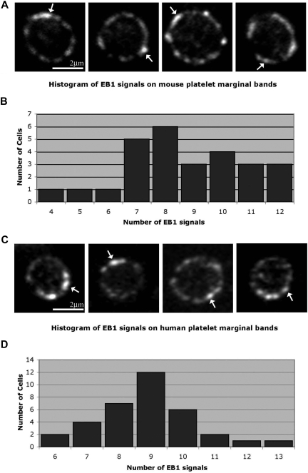
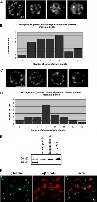
 ) are labeled along the marginal band. An average of 8.66 (± 2.11; n = 26) comets were observed along the marginal band. (B) Histogram showing EB1 signals observed along the microtubule coil of each mouse platelet. The number of signals ranged from 4 to 12, with a median of 8. (C) Anti-EB1 staining in resting human platelets. EB1 comets () are identified. An average of 8.9 (± 1.5; n = 34) comets are seen along each coil. (D) Histogram of EB1 signals along the microtubule coil of human platelets. The number of signals ranged from 6 to 13, with a median of 9.
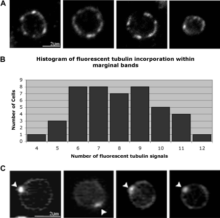
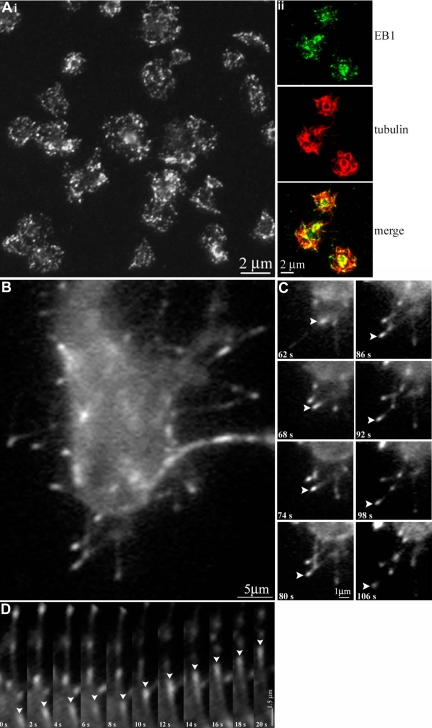
 ) along the platelet marginal band.
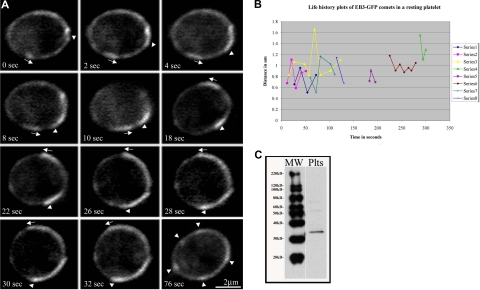 ) and counterclockwise () over the course of 32 seconds. The final panel (76 seconds) shows the same cell with several EB3-GFP comets and foci (). (B) Life history plots of 8 individual EB3-GFP comets within a resting platelet show distance traveled over time. (C) Western blot detection of endogenously expressed EB3 in platelet lysates.
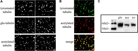
Comment in
-
Platelet marginal bands: not so marginal.Blood. 2008 May 1;111(9):4423. doi: 10.1182/blood-2008-02-137786. Blood. 2008. PMID: 18441237 No abstract available.
References
-
- Behnke O. Microtubules in disk-shaped blood cells. Int Rev Exp Pathol. 1970;9:1–92. - PubMed
-
- Kenney DM, Linck RW. The cytoskeleton of unstimulated blood platelets: structure and composition of the isolated marginal microtubular band. J Cell Sci. 1985;78:1–22. - PubMed
-
- Lewis SA, Gu W, Cowan N. Free intermingling of mammalian Beta-tubulin isotypes among functionally distinct microtubules. Cell. 1987;49:539–548. - PubMed
Publication types
MeSH terms
Grants and funding
LinkOut - more resources
Full Text Sources
Molecular Biology Databases
