. 2008 Jan 30;3(1):e1527.
doi: 10.1371/journal.pone.0001527.

Anti-human tissue factor antibody ameliorated intestinal ischemia reperfusion-induced acute lung injury in human tissue factor knock-in mice

Affiliations

Anti-human tissue factor antibody ameliorated intestinal ischemia reperfusion-induced acute lung injury in human tissue factor knock-in mice

Xiaolin He et al. PLoS One. .

Abstract

Background: Interaction between the coagulation and inflammation systems plays an important role in the development of acute respiratory distress syndrome (ARDS). Anti-coagulation is an attractive option for ARDS treatment, and this has promoted development of new antibodies. However, preclinical trials for these antibodies are often limited by the high cost and availability of non-human primates. In the present study, we developed a novel alternative method to test the role of a humanized anti-tissue factor mAb in acute lung injury with transgenic mice.

Methodology/principal findings: Human tissue factor knock-in (hTF-KI) transgenic mice and a novel humanized anti-human tissue factor mAb (anti-hTF mAb, CNTO859) were developed. The hTF-KI mice showed a normal and functional expression of hTF. The anti-hTF mAb specifically blocked the pro-coagulation activity of brain extracts from the hTF-KI mice and human, but not from wild type mice. An extrapulmonary ARDS model was used by intestinal ischemia-reperfusion. Significant lung tissue damage in hTF-KI mice was observed after 2 h reperfusion. Administration of CNTO859 (5 mg/kg, i.v.) attenuated the severity of lung tissue injury, decreased the total cell counts and protein concentration in bronchoalveolar lavage fluid, and reduced Evans blue leakage. In addition, the treatment significantly reduced alveolar fibrin deposition, and decreased tissue factor and plasminogen activator inhibitor-1 activity in the serum. This treatment also down-regulated cytokine expression and reduced cell death in the lung.

Conclusions: This novel anti-hTF antibody showed beneficial effects on intestinal ischemia-reperfusion induced acute lung injury, which merits further investigation for clinical usage. In addition, the use of knock-in transgenic mice to test the efficacy of antibodies against human-specific proteins is a novel strategy for preclinical studies.


Conflict of interest statement

Competing Interests: The authors have declared that no competing interests exist.

Figures

Figure 1
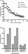
Figure 1. Human tissue factor is functionally expressed in hTF-KI transgenic mice, and effectively and specifically inhibited by the anti-hTF mAb, CNTO859.
Pro-coagulant activity of TF in brain extracts from either hTF-KI or wild type (WT) mice, or from human brain tissue was measured with a one-stage clotting assay, and a similar prothrombin time was seen in all brain extracts, indicating a functional replacement of mTF by hTF expressed in the hTF-KI mice (A). Anti-hTF antibody, CNTO859, dose-dependently inhibited TF pro-coagulant activity in the brain extracts from hTF-KI mice and human, but not wild type mice (B). The experiments were repeated three times, and representative data from one experiment are shown.
Figure 2
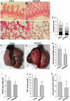
Figure 2. Administration of anti-hTF mAb ameliorated intestinal ischemia-reperfusion (IIR)-induced acute lung injury in hTF-KI mice.
IIR challenged hTF-KI mice were treated with CNTO859 (5 mg/kg, i.v.) or saline. The representative histology (H&E, x400) from the intestine (A, B) and lung (C, D) was shown. The lung injury was scored by a pathologist in a blind fashion (E) (▪ inflammatory cells infiltration; ▪ alveolar wall edema; ▪ hemorrhage; atelectasis). □ Lung injury scores of 4 categories were analyzed with Kruskall-Wallis test, n = 4 animals/group, *: p<0.05. The pulmonary permeability was determined by Evans Blue dye assay (F, G, H). Administration of CNTO859 also reduced the wet/dry lung weight ratio (I), albumin concentration (J), and total cell counts (K) in the BAL fluid. Panels I-K: n = 4 animals/group,*: p<0.05, un-paired t-test.
Figure 3
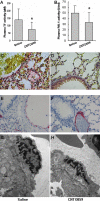
Figure 3. Anti-hTF mAb treatment attenuated IIR induced coagulapathy and protected pulmonary endothelium.
Plasma TF and PAI-1 activities were determined as described in Methods. Administration of CNTO859 significantly inhibited both TF (A) and PAI-1 (B) activities in comparison with the saline treated control (*: p<0.05, n = 4 animals/group, un-paired t-test). The fibrin staining showed that CNTO859 ameliorated IIR-induced fibrin deposition (pink) in the alveoli (C, D). The lung tissues were stained for vWF, a specific marker for endothelial integrity. Weaker staining of vWF (pink) of endothelium in the pulmonary vessels was noted in the saline control group (E). In CNTO859 treated animals stronger vWF staining was observed in the endothelium of pulmonary vessels (F). The pulmonary endothelial injury was further examined with electron microscopy. IIR challenge led to significant endothelial cell swelling (G), which was protected by CNTO859 treatment (H).
Figure 4
Figure 4. Anti-hTF mAb treatment reduced inflammatory response.
Inflammatory cytokines were measured in the lung tissues and BAL fluid with a cytometric bead array. The expression levels of IL-6 (A), TNFα (B), and MCP-1 (C) in the lung tissues were significantly reduced by CNTO859 in comparison to saline group. In the BAL fluid CNTO859 also reduced the IL-6 levels (D), but the IL-10 level remained unchanged between the two groups (E). Un-paired t-test was used, *: p<0.05, n = 4 animals/group.
Figure 5
Figure 5. Anti-hTF mAb treatment reduced cell death.
IIR-induced cell death in the lungs was determined by TUNEL staining. CNTO859 effect on the cell death was shown in representative slides (400x) (A, B), and quantified by counting the TUNEL positive cells from 10 randomly chosen fields (C). Caspase 3 activity in the lung tissue was also reduced by the anti-hTF antibody treatment (D). Un-paired t-test was used, 4 animals per group, *: p<0.05.

References

    1. Liu M. Searching for acute respiratory distress syndrome genes: aren't we there yet? Am J Respir Crit Care Med. 2005;171:298–299. - PubMed
    1. Rijneveld AW, Weijer S, Bresser P, Florquin S, Vlasuk GP, et al. Local activation of the tissue factor-factor VIIa pathway in patients with pneumonia and the effect of inhibition of this pathway in murine pneumococcal pneumonia. Crit Care Med. 2006;34:1725–1730. - PubMed
    1. Abraham E. Coagulation abnormalities in acute lung injury and sepsis. Am J Respir Cell Mol Biol. 2000;22:401–404. - PubMed
    1. Carraway MS, Welty-Wolf KE, Miller DL, Ortel TL, Idell S, et al. Blockade of tissue factor: treatment for organ injury in established sepsis. Am J Respir Crit Care Med. 2003;167:1200–1209. - PubMed
    1. Schultz MJ, Millo J, Levi M, Hack CE, Weverling GJ, et al. Local activation of coagulation and inhibition of fibrinolysis in the lung during ventilator associated pneumonia. Thorax. 2004;59:130–135. - PMC - PubMed

Publication types

MeSH terms

LinkOut - more resources