The homeodomain transcription factor Cdx1 does not behave as an oncogene in normal mouse intestine
- PMID: 18231635
- PMCID: PMC2213897
- DOI: 10.1593/neo.07703
The homeodomain transcription factor Cdx1 does not behave as an oncogene in normal mouse intestine
Abstract
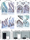
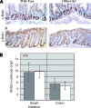
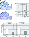
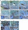
The Caudal-related homeobox genes Cdx1 and Cdx2 are intestine-specific transcription factors that regulate differentiation of intestinal cell types. Previously, we have shown Cdx1 to be antiproliferative and to promote cell differentiation. However, other studies have suggested that Cdx1 may be an oncogene. To test for oncogenic behavior, we used the murine villin promoter to ectopically express Cdx1 in the small intestinal villi and colonic surface epithelium. No changes in intestinal architecture, cell differentiation, or lineage selection were observed with expression of the transgene. Classic oncogenes enhance proliferation and induce tumors when ectopically expressed. However, the Cdx1 transgene neither altered intestinal proliferation nor induced spontaneous intestinal tumors. In a murine model for colitis-associated cancer, the Cdx1 transgene decreased, rather than increased, the number of adenomas that developed. In the polyps, the expression of the endogenous and the transgenic Cdx1 proteins was largely absent, whereas endogenous Villin expression was retained. This suggests that transgene silencing was specific and not due to a general Villin inactivation. In conclusion, neither the ectopic expression of Cdx1 was associated with changes in intestinal cell proliferation or differentiation nor was there increased intestinal cancer susceptibility. Our results therefore suggest that Cdx1 is not an oncogene in normal intestinal epithelium.
Figures


Similar articles
-
Cdx1 inhibits human colon cancer cell proliferation by reducing beta-catenin/T-cell factor transcriptional activity.J Biol Chem. 2004 Aug 27;279(35):36865-75. doi: 10.1074/jbc.M405213200. Epub 2004 Jun 23. J Biol Chem. 2004. PMID: 15215241
-
Transgenic Cdx2 induces endogenous Cdx1 in intestinal metaplasia of Cdx2-transgenic mouse stomach.FEBS J. 2009 Oct;276(20):5821-31. doi: 10.1111/j.1742-4658.2009.07263.x. Epub 2009 Sep 2. FEBS J. 2009. PMID: 19725873
-
CDX transcription factors positively regulate expression of solute carrier family 5, member 8 in the colonic epithelium.Gastroenterology. 2010 Feb;138(2):627-35. doi: 10.1053/j.gastro.2009.10.047. Epub 2009 Nov 10. Gastroenterology. 2010. PMID: 19900445
-
The role of Cdx proteins in intestinal development and cancer.Cancer Biol Ther. 2004 Jul;3(7):593-601. doi: 10.4161/cbt.3.7.913. Epub 2004 Jul 9. Cancer Biol Ther. 2004. PMID: 15136761 Review.
-
YAP oncogene overexpression supercharges colon cancer proliferation.Cell Cycle. 2012 Mar 15;11(6):1090-6. doi: 10.4161/cc.11.6.19453. Epub 2012 Mar 15. Cell Cycle. 2012. PMID: 22356765 Free PMC article. Review.
Cited by
-
Dinosaurs and ancient civilizations: reflections on the treatment of cancer.Neoplasia. 2010 Dec;12(12):957-68. doi: 10.1593/neo.101588. Neoplasia. 2010. PMID: 21170260 Free PMC article.
-
Cdx genes, inflammation, and the pathogenesis of intestinal metaplasia.Prog Mol Biol Transl Sci. 2010;96:231-70. doi: 10.1016/B978-0-12-381280-3.00010-5. Prog Mol Biol Transl Sci. 2010. PMID: 21075347 Free PMC article. Review.
-
Transcription Factor SP2 Enhanced the Expression of Cd14 in Colitis-Susceptible C3H/HeJBir.PLoS One. 2016 May 18;11(5):e0155821. doi: 10.1371/journal.pone.0155821. eCollection 2016. PLoS One. 2016. PMID: 27191968 Free PMC article.
-
Methylation-dependent activation of CDX1 through NF-κB: a link from inflammation to intestinal metaplasia in the human stomach.Am J Pathol. 2012 Aug;181(2):487-98. doi: 10.1016/j.ajpath.2012.04.028. Epub 2012 Jun 27. Am J Pathol. 2012. PMID: 22749770 Free PMC article.
-
CDX1 confers intestinal phenotype on gastric epithelial cells via induction of stemness-associated reprogramming factors SALL4 and KLF5.Proc Natl Acad Sci U S A. 2012 Dec 11;109(50):20584-9. doi: 10.1073/pnas.1208651109. Epub 2012 Oct 29. Proc Natl Acad Sci U S A. 2012. PMID: 23112162 Free PMC article.
References
-
- Crosnier C, Stamataki D, Lewis J. Organizing cell renewal in the intestine: stem cells, signals and combinatorial control. Nat Rev Genet. 2006;7:349–359. - PubMed
-
- van Es JH, Jay P, Gregorieff A, van Gijn ME, Jonkheer S, Hatzis P, Thiele A, van den Born M, Begthel H, Brabletz T, et al. Wnt signalling induces maturation of paneth cells in intestinal crypts. Nat Cell Biol. 2005;7:381–386. - PubMed
-
- Yang Q, Bermingham NA, Finegold MJ, Zoghbi HY. Requirement of Math1 for secretory cell lineage commitment in the mouse intestine. Science. 2001;294:2155–2158. - PubMed
-
- Fre S, Huyghe M, Mourikis P, Robine S, Louvard D, Artavanis-Tsakonas S. Notch signals control the fate of immature progenitor cells in the intestine. Nature. 2005;435:964–968. - PubMed
Publication types
MeSH terms
Substances
Grants and funding
LinkOut - more resources
Full Text Sources
Molecular Biology Databases
Research Materials
