. 2008 Jan;10(1):41-51.
doi: 10.1593/neo.07754.

Emodin-induced generation of reactive oxygen species inhibits RhoA activation to sensitize gastric carcinoma cells to anoikis

Affiliations

Emodin-induced generation of reactive oxygen species inhibits RhoA activation to sensitize gastric carcinoma cells to anoikis

Jun Cai et al. Neoplasia. 2008 Jan.

Abstract

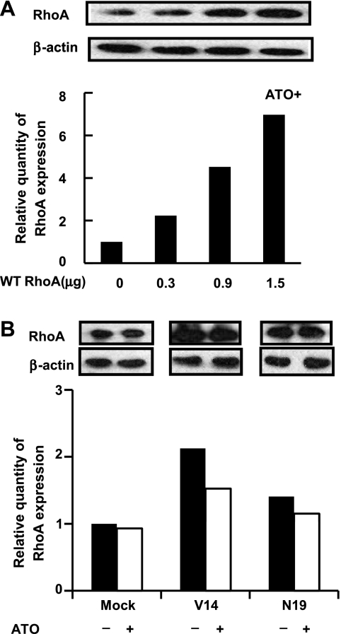
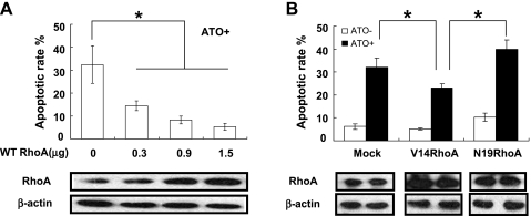
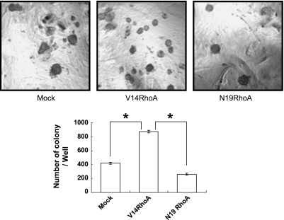
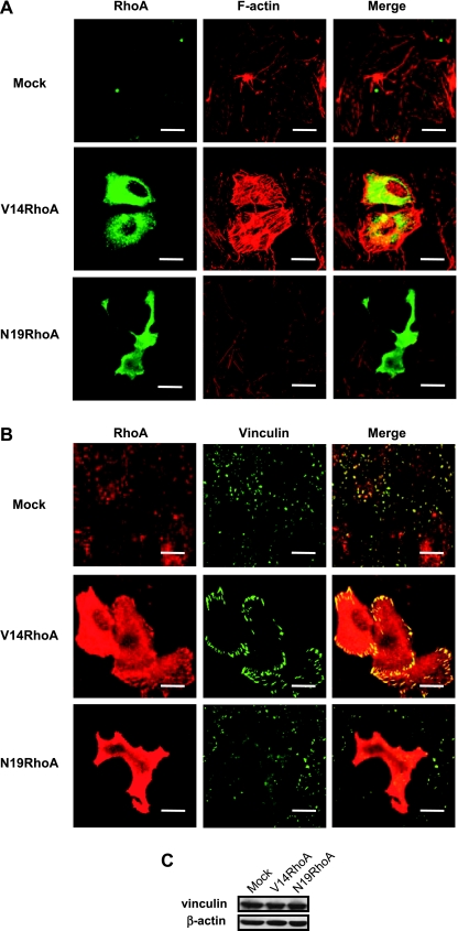
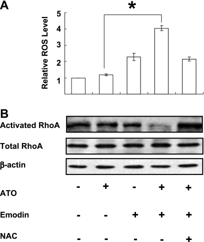
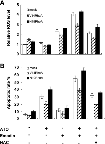
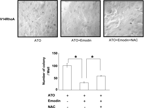
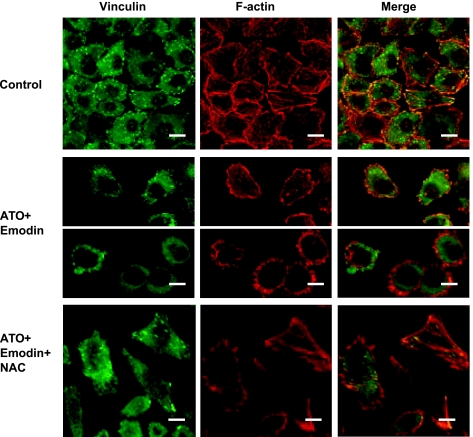
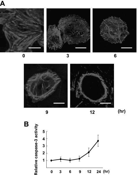
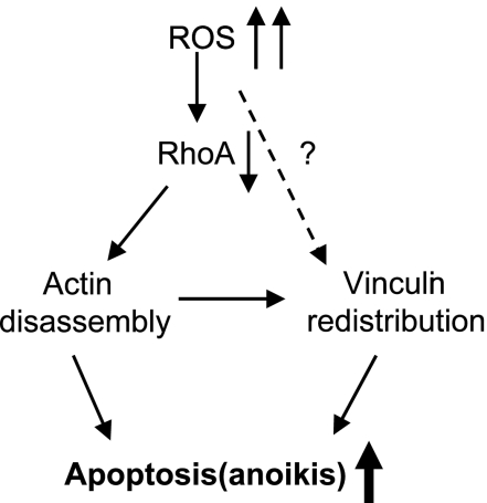
RhoA is a critical signaling molecule regulating a variety of cellular processes, such as cytoskeletal organization, adhesion, and apoptosis. It is recently considered responsive to reactive oxygen species (ROS). Nevertheless, how RhoA regulates anoikis, a detachment-initiated apoptosis, and how this regulation is affected by ROS are not clear. The present study investigated the role of RhoA in apoptosis/anoikis in gastric cancer cells and the changes of RhoA and anoikis under oxidative stress. Immunohistochemistry showed that RhoA expression was upregulated in the primary gastric carcinoma compared with normal gastric mucosa. Overactivation of RhoA by transfection with the V14RhoA mutant prevented gastric cancer line SGC-7901 cells from arsenic-induced apoptosis and conferred anoikis resistance through, at least in part, promoting formations of F-actin fibers and focal adhesion. Oxidative stress caused by emodin, an ROS producer, in combination with arsenic trioxide (ATO) led to RhoA inactivation that triggered structural disruption of focal adhesion complex and eventually resulted in anoikis, and these effects could be partially reversed by antioxidant N-acetylcysteine (NAC). In conclusion, activation of RhoA is required for the maintenance of anoikis resistance phenotype of gastric cancer cells, and oxidative stress might be a therapeutic strategy for the inhibition of RhoA in cancer cells.


Figures

Figure 1
Figure 1
RhoA was overexpressed in gastric carcinoma tissues, and the level of expression was related to malignancy (peroxidase-DAB immunohistochemistry): (A) normal gastric mucosa, (B) highly differentiated gastric carcinoma, (C) moderately differentiated gastric carcinoma, and (D) lowly differentiated gastric carcinoma (original magnification, x200).
Figure 2
Figure 2
Overexpression or overactivation of RhoA in SGC-7901 cells antagonized ATO-induced apoptosis (Annexin V/PI flow cytometry). (A) Cells were transfected with wild-typed (WT) RhoA at increased doses and were exposed to ATO (5 µM) for 48 hours. (B) Cells were transfected with mock DNA, V14RhoA, and N19RhoA at 40 hours before treatment with or without ATO (5 µM) for 48 hours. The quantity of RhoA expression was controlled by Western blot analysis. Values were mean ± SD of three different experiments. ANOVA was used for comparison of each group. *P < .05.
Figure 3
Figure 3
RhoA activation promoted anoikis resistance of SGC-7901 cells (colony formation in soft agar). Cells were seeded in the agar at 40 hours posttransfection and remained growing for 2 weeks before colony calculation (original magnification, x50). Values were mean ± SD of three different experiments. ANOVA was used for comparison of each group. *P < .05.
Figure 4
Figure 4
RhoA activation changed the assembly of actin and distribution of vinculin (confocal microscopy and Western blot analysis). Cells were assayed at 40 hours posttransfection with the vector DNA or the RhoA mutants. (A) RhoA immunofluorescence (green) and F-actin staining with rhodamine-phalloidin (red). (B) RhoA and vinculin double immunofluorescence (red for RhoA and green for vinculin). (C) Western blot analysis for vinculin and actin. Scale bar, 10 µm.
Figure 5
Figure 5
Oxidative stress caused by emodin in combination with arsenic suppressed the activation of RhoA, but did not downregulate the expression of total RhoA (DCF flow cytometry, GST-RBD pull-down, and Western blot). (A) Relative cellular ROS level of native SGC-7901 cells after 1-hour treatments. ATO, 5 µM; Emodin, 10 µM; NAC, 10 mM. Values were mean ± SD of three different experiments. ANOVA was applied for comparison of the means of two groups. *P < .05. (B) Western blot for RhoA of native SGC-7901 cells after 9-hour treatments. ATO, 5 µM; Emodin, 10 µM; NAC, 10 mM. Blots were representative of two experiments.
Figure 6
Figure 6
Oxidative stress caused by emodin in combination with arsenic-enhanced apoptosis, and these effects could be partially reversed by NAC. (A) Relative ROS level after 1-hour treatments in SGC-7901 cells 40 hours posttransfection with DNA for mock and RhoA mutants: ATO, 5 µM; Emodin, 10 µM; NAC, 10 mM. ATO versus ATO + Emodin: P < .05. (B) Apoptotic rates after 48-hour treatments in cells with transfections and treatments similar to panel A. ATO versus ATO + Emodin: P < .05; ATO + Emodin versus ATO + Emodin + NAC: P < .05. Values were mean ± SD of three different experiments. ANOVA was used for comparison of each group.
Figure 7
Figure 7
Oxidative stress caused by emodin in combination with arsenic inhibited anoikis resistance of SGC-7901 cells transfected with V14RhoA (colony formation in soft agar). Cells were seeded in the agar after drug treatments for 9 hours at 40 hours posttransfection of V14RhoA, and remained growing for 2 weeks before calculation (original magnification, x50). ATO, 5µM; Emodin, 10µM; NAC, 10 mM. Values were mean ± SD of three different experiments. ANOVA was applied for comparison of each group. *P < .05.
Figure 8
Figure 8
Oxidative stress caused by emodin in combination with arsenic-altered assembly of F-actin and distribution of vinculin, especially in apoptotic cells, whereas NAC could antagonize this effect (confocal microscopy). Native SGC-7901 cells were exposed to drugs for 12 hours. ATO, 5 µM; Emodin, 10 µM; NAC, 10 mM. Scale bar, 10 µm. Two parts of the middle panel showed cells separated in the field with two typical morphologies of cell detachment. Cells in the upper parts of the middle panel started to detach and the lower parts of the middle panel were round up with cortex actin rings.
Figure 9
Figure 9
Oxidative stress caused by emodin in combination with arsenic-induced disassembly of F-actin that preceded caspase-3 activation. (A) Confocal microscopy showing time course of Factin disassembly of native SGC-7901 cells after the combinative treatment (ATO + Emodin: ATO, 5 µM; Emodin, 10 µM; scale bar, 10 µm). (B) Time course for relative caspase-3 activities of native SGC-7901 cells after the same treatment as in A. Values were mean ± SD of three different experiments.
Figure 10
Figure 10
Hypothetical model for the role of RhoA in ROS-induced apoptosis.

Similar articles

Cited by

References

    1. Hall A. RhoA GTPases and the actin cytoskeleton. Science. 1998;279:509–514. - PubMed
    1. Ridley AJ. Rho family proteins: coordinating cell responses. Cancer Metastasis Rev. 2001;11:471–477. - PubMed
    1. Coleman ML, Olson MF. Rho GTPase signaling pathways in the morphological changes associated with apoptosis. Cell Death Differ. 2002;9:493–504. - PubMed
    1. Etienne-Manneville S, Hall A. Rho GTPases in cell biology. Nature. 2002;420:629–635. - PubMed
    1. Vega FM, Ridley AJ. SnapShot: Rho Family GTPases. Cell. 2007;129:1430–1431. - PubMed

Publication types

MeSH terms